Download
1 / 12
140 likes | 408 Vues
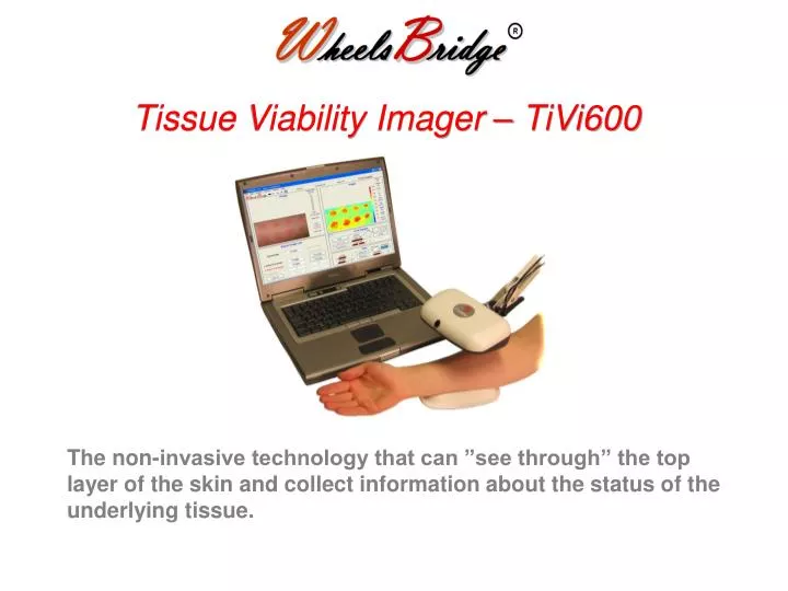
Tissue Viability Imager – TiVi600. The non-invasive technology that can ”see through” the top layer of the skin and collect information about the status of the underlying tissue. Skin testing. Drug development. Development of skin care products and cosmetics Microvascular research.

E N D
Tissue Viability Imager – TiVi600 The non-invasive technology that can ”see through” the top layer of the skin and collect information about the status of the underlying tissue.
Skin testing Drug development Development of skin care products and cosmetics Microvascular research Applications
flash detector polarization photo filter secondfilter blocks polarized light green plane red plane polarized light depola-rized light algorithm skin tissue viability image Principle Polarization Spectroscopy red light is moderately absorbed by RBCs green light is highly absorbed by RBCs
A transparent tubing system filled with RBCs of different concentration is used for modelling the skin microcirculation. Simulated and measured TiVi-index versus RBC fraction A linear relationship is attained between RBC concentration and TiVi output signal. Validation Digital camera
Features Small and portable design. 12 images per minute. 2D color presentation. Video-clip presentation. Basic data availability. Benefits Non-invasive and remote operation. Instantaneous image capturing. Not sensitive to movements or ambient light. User friendly. Does not influnce the microcirculation. Calibration not necessary.
Before application of methyl nicotinate. About ten minutes following application of methyl nicotinate. Select regions of interest. Display the Erythema intensity versus time curves. Analysis of time series Build a video out of 100 images.
Select a cross-section. Display the TiVi values along the cross-section versus time. Analysis of time series
Apply topically: 0.5 mg/ml clobestal proprionate (20 microliter). 50 mmol methyl nicotinate (20 microliter). The average TiVi-values for the erythema, blanching and control area. Erythema and Blanching
Step 1. Drug application by iontophoresis. Step 2. Capture a sequence of images through the ring-shape iontophoresis electrode and watch the result. Step 3. Display the time-sequence of average TiVi-values. Step 4. Generate a dose-response curve. Transcutaneous drug delivery
Comparison: Tissue Viability Imaging Laser Doppler Perfusion Imaging TiVi Quantity LDPI RBC conc Measurand RBC vel * conc none Movement artifact sensitivity high up to 9.000.000 No. ofmeasurement points up to 65.000 low Ambient light sensitivity high instantaneous Image capturing time minutes good Portability poor
Thank You! more info: www.wheelsbridge.se