Download
1 / 30
320 likes | 488 Vues
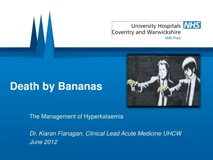
Death by Bananas. The Management of Hyperkalaemia Dr. Kiaran Flanagan, Clinical Lead Acute Medicine UHCW June 2012. Case 1. Patient comes into ED referred by GP for high potassium of 6.7 You see the notes in the SIFT tray What do you do?. How to manage.

E N D
Death by Bananas The Management of Hyperkalaemia Dr. Kiaran Flanagan, Clinical Lead Acute Medicine UHCW June 2012
Case 1 • Patient comes into ED referred by GP for high potassium of 6.7 • You see the notes in the SIFT tray • What do you do? ...
How to manage • Pick up notes and PUT STICKER ON THE LIST • ABCDE • What are you likely to find? • What urgent investigation do you need to make a treatment decision? • What action would you take if • 1. Normal • 2. Abnormal
What next? • History... • Examination... • Further tests • What are you looking for?
What next... • Senior review? • Actions you should recommend... • Drugs • Monitoring • Admit/ Discharge • Further checks • Anticipated future actions • How will you make this happen?
Case 2 • Patient on the ward • ATSP – unwell, vomiting • Day 2 of admission – post op R hemicolectomy • What do you do?
What do you do? • ABCDE • Investigations? • Monitoring...
Patient hyperkalaemic • What else do you look for? • What test needs to have been done? • What treatment do you need to give?
Recheck K at 3 hours • Still high... • What next? • Treatment • Advice • Monitoring
Recheck K at 6 hours • Still high... • What do you do? • Treatment • Monitoring • Ask for help • Who • What will you tell them and how? • What for
Case 3 • Patient – Medical ALERT to Resus • Drowsy • High glucose • What do you do????
What do you do? • ABCDE • Urgent tests • What is the diagnosis? • How do you manage?
Case 4 • Cardiac Arrest Call • PEA • What do you think about?
Hyperkalaemia in cardiac arrest • What do you give?
Case 5 • Called to ward 1, pt unwell • Low BP, low glucose, high potassium • What do you do? • Assessment • Further tests? • Working diagnosis • Treatment?
Causes of Hyperkalaemia • Decreased or impaired potassium excretion – renal failure, potassium-sparing diuretics, urinary obstruction, sickle cell disease, Addison disease, and systemic lupus erythematosus (SLE) • Additions of potassium into extracellular space - potassium supplements (eg, PO/IV potassium, salt substitutes), rhabdomyolysis, and hemolysis (eg, blood transfusions, burns, tumor lysis) • Transmembrane shifts (ie, shifting potassium from the intracellular to extracellular space) - acidosis and medication effects (eg, acute digitalis toxicity, beta-blockers, succinylcholine) • Factitious or pseudohyperkalemia - improper blood collection (eg, ischemic blood draw from venipuncture technique), laboratory error, leukocytosis, and thrombocytosis
Causes • Ineffective elimination • Kidneys • Drugs • Endocrine • Excessive release from cells • Injury • Metabolic • Excessive intake • Lethal Injection • Pseudo
How does it affect the heart? • Hyperkalemia results in: • Inhibition of atrial myocardial depolarization. • Slowing of heart rate. • Prolonging QRS duration; complexes may become bizarre. • Also known as atrial standstill. • Rhythm called sinoventricular rhythm. • The ECG is a poor substitute for serum potassium levels to determine the degree of abnormality
ECG Changes • From reduction of P wave amplitude and prolongation of PR interval to absence of P waves altogether. • Increase of QRS duration. • Increase of QT duration. • Slowing of heart rate. • T waves become tall and spiked. • Decreased R wave amplitude
3 Principles of Treatment • Stabilise myocardium • Move it into cells • Increase elimination
Dextrose - Insulin • How does it work? • How long for? • How do you give it? • What is the dose? • Other considerations...
Calcium Gluconate • How does it work? • How long for? • How do you give it? • What is the dose? • Other considerations...
Sodium Bicarbonate • How does it work? • How long for? • How do you give it? • What is the dose? • Other considerations...
Calcium Resonium • Hmmm....
More controversial • Salbutamol • Furosemide
Protocols • If K > 6 mmol • Calcium Resonium • Unless – Rising fast/ patient septic then treat as below • If K > 6.5 – normal ECG • Dextrose Insulin • Calcium Resonium
Protocols • If K > 6.5 – abnormal ECG or • If K > 7 • Calcium Gluconate • Dex Insulin • Salbutamol • Sodium Bicarbonate • RRT
Important Bits... • POTENTIAL LIFE THREATENING EMERGENCY • TREAT IF INDICATED • TRUST BUT VERIFY • RECHECK • CARDIAC MONITORING • EXPERT HELP • PREVENTION