Download
1 / 110
1.1k likes | 1.29k Vues
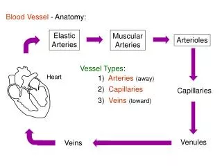
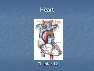
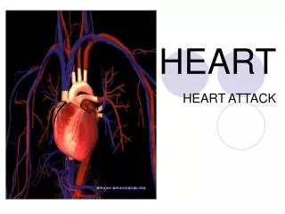
Heart . Congestive heart failure. or heart failure : condition : heart is unable to adequately pump blood throughout the body. Characterized : shortness of breath (dyspnea) abnormal fluid retention, which usually results in swelling (edema) in the feet and legs. . Heart failure.

E N D
Congestive heart failure • or heart failure : condition : heart is unable to adequately pump blood throughout the body
Characterized : • shortness of breath (dyspnea) • abnormal fluid retention, which usually results in swelling (edema) in the feet and legs.
Heart failure • Left-sided heart failure • Right-sided heart failure
Left-sided heart failure. When LVcannot adequately pump blood out of the left atrium, or when one or more of the heart valves becomes leaky or narrowed (stenotic), blood can "back up" into the lungs
left-sided heart failure: • lungs become congested with fluid (called pulmonary edema), • causing difficulty breathing and • interfering with the movement of oxygen from the lungs into the bloodstream, causing fatigue.
Right-sided heart failure • abnormality or condition affects the flow of blood through the right ventricle, pressure in the blood vessels increases and fluid is forced from the blood vessels into body tissues. • causes swelling (edema), usually in the feet and legs, and sometimes, in the abdomen.
The NYHA functional class (the New York Heart Association) • determine how much CHF limits their lifestyle • Useful in following the course of disease and assessing the effects of therapy • Aid in the dental management
Class I: No symptoms at any level of exertion, no limitation of physical activity Class II: Slight limitation of physical activity. Fatigue, palpitations and dyspnea with ordinary physical activity but comfortable at rest Class III: marked limitation of activity. Less than ordinary physical activity results in symptoms, but patients are comfortable at rest Class IV: Symptoms are present at rest, and any physical exertion exacerbates the symptoms
Congestive heart failure Potential problem related to dental care 1. sudden death from cardiac arrest or arrhythmia 2. Myocardial infarction 3. CVA 4. Infective endocarditis if CHF is caused by rheumatic heart dis., congenital heart dis.
CHF Potential problem related to dental care 5. Shortness of breath 6. Drug side effects : orthostatic hypotension (diuretics,vasodilators) arrhythmia (digoxin overdose) nausea, vomiting (digoxin, vasodilators) palpitations (vasodilators) 7. Infection
Prevention of complication 1. Detection and referral to physician 2. No routine dental care until under good medical management (class I or II and possibly III) 3. Good medical management – cause of heart failure - hypertension - valvular dis. (rheumatic heart dis.) - congenital heart dis., MI - Renal failure - Thyrotoxicosis - chronic obstructive lung disease
4. Class I or II, use max. 0.036 mg epinephrine avoid vasoconstrictors in class III or IV 5. Semisupine or upright position (decrease collection of fluid in lung) 6. Terminate appointment if patient becomes fatigue 7. Drug considerations digitalis – N/V anticoagulants - PT = 2times or less, - INR = 3.0 or less antidysrhythmic agents, antihypertensive avoidance of outpatient general anesthesia
CHF Emergency care 1. Conservative in acute congestive failure: drug for pain control and antibiotics for infection 2. Under good medical management: deal with underlying cause and presence of any complications in dental management
Endocarditis • Inflammation of endocardium • most common structures involved are the heart valves. • Endocarditis can be classified by etiology as either infective or non-infective
Infective endocarditis • valves of the heart do not actually receive any blood supply of their own, defense mechanisms (such as white blood cells) cannot enter. • If an organism (such as bacteria) hold on the valves, the body cannot get rid of them.
If valve damaged (for instance in rheumatic fever) bacteria have a chance to hold. • clinically divided into • acute and subacute endocarditis. This classifies both the tempo of progression and severity of disease.
Subacute bacterial endocarditis (SBE) : often due to streptococci of low virulence and mild to moderate illness which progresses slowly over weeks and months • Acute bacterial endocarditis (ABE) : fulminant illness over days to weeks, more likely due to Staphylococcus aureus (greater virulence, or disease-producing capacity)
Aetiology and pathogenesis • altered blood flow around the valves is a risk factor in obtaining endocarditis. • The valves may be damaged congenitally, from surgery, by auto-immune mechanisms, or simply as a consequence of old age. • The damaged part of a heart valve becomes covered with a blood clot, a condition known as non-bacterial thrombotic endocarditis (NBTE).
In healthy individual, a bacteraemia would normally be cleared quickly with no adverse consequences. • If a heart valve is damaged and covered with a piece of a blood clot, the valve provides a place for the bacteria to attach themselves and an infection can be established.
The bacteraemia is often caused by minor dental procedures, such as a tooth removal. • Another causes result from a high number of bacteria getting into the bloodstream. (Colorectal cancer, serious urinary tract infections and IV drug use) • With a large number of bacteria, even a normal heart valve may be infected. • A more virulent organism (Staphylococcus aureus) is usually responsible for infecting a normal valve.
Intravenous drug users : right heart valves infected (veins that are injected enter the right side of the heart) • The injured valve is most commonly affected when there is a pre-existing disease. (rheumatic heart disease this is the aortic and the mitral valves) : left heart valves
Clinical and pathological features • Fever (often spiking) • Continuous presence of micro-organisms in the bloodstream determined by serial collection of blood cultures • Vegetations on valves on echocardiography • Septic emboli, causing circulatory problems (stroke, gangrene of fingers) • Chronic renal failure
Clinical and pathological features • Osler's nodes (painful subcutaneous lesions in the distal fingers) • Janeway lesions (painless hemorrhagic cutaneous lesions on the palms and soles) • Roth spots on the retina • Conjunctival petechiae • A new or changing heart murmur, particularly murmurs suggestive of valvular incompetence • Splinter haemorrhages
Micro-organisms responsible • Many types of organism • isolated by blood culture • Alpha-haemolytic streptococci, that are present in the mouth will often be the organism isolated if a dental procedure caused the bacteraemia.
If bacteraemia was introduced through the skin, such as contamination in surgery, during catheterisation, or in an IV drug user Staphylococcus aureus
A third important cause of endocarditis is Enterococci(abnormalities in the gastrointestinal or urinary tracts) • Enterococci : causes of nosocomial or hospital-acquired endocarditis. • alpha-haemolytic streptococci and Staphylococcus aureus : causes of community-acquired endocarditis.
Treatment • High dose antibiotics ( by intravenous route) • Antibiotics are continued for a long time, typically two to six weeks. • Surgical removal of the valve is necessary in patients who fail to clear micro-organisms from their blood in response to antibiotic therapy, or in patients who develop cardiac failure resulting from destruction of a valve
A removed valve is usually replaced with an artificial valve which may either be mechanical (metallic) or obtained from an animal such as a pig (bioprosthetic valves) • Infective endocarditis is associated with a 25% mortality.
ENDOCARDITIS RISK STRATIFICATION Endocarditis prophylaxis recommended High risk • Prosthetic heart valves • Prior bacterial endocarditis • Complex cyanotic congenital heart disease • Surgically constructed systemic pulmonary shunts or conduct
ENDOCARDITIS RISK STRATIFICATION Endocarditis prophylaxis recommended Moderate risk • Most other congenital cardiac malformations • Acquired valvular dysfunction • Hypertrophic cardiomyopathy • Mitral valve prolapse with regurgitations and/or thickened leaflets
ENDOCARDITIS RISK STRATIFICATION Endocarditis prophylaxis not recommended Negligible risk • Isolated secundum atrial septal defect • Surgical repair of ASD, VSD, or PDA (without residua beyond 6 mo.) • Prior coronary artery bypass graft • Mitral valve prolapse without regurgitation • Physiologic, functional, or innocent heart murmurs
ENDOCARDITIS RISK STRATIFICATION Endocarditis prophylaxis not recommended Negligible risk • Previous Kawasaki dis. without valvular dysfunction • Previous rheumatic fever without valvular dysfunction • Cardiac pacemakers and implanted defibrillators
DENTAL AND ORAL SURGERY PROCEDURES AND ENDOCARDITIS PROPHYLAXIS Endocarditis prophylaxis recommended in high- and moderate-risk cardiac conditions • Exodontia • Periodontal procedures • Incision and drainage of abscesses • Dental implant placement and uncovering • Reimplantation of avulsed teeth • Endodontic therapy or apical surgery • Placement of intermaxillary fixation
DENTAL AND ORAL SURGERY PROCEDURES AND ENDOCARDITIS PROPHYLAXIS Endocarditis prophylaxis recommended in high- and moderate-risk cardiac conditions • Reduction of contaminated maxillofacial fractures • Osteotomies • Subgingival placement of antibiotic fibers or strips • Intraligamentary LA injections • Prophylactic dental or implant cleaning • Intraoral biopsies
DENTAL AND ORAL SURGERY PROCEDURES AND ENDOCARDITIS PROPHYLAXIS Endocarditis prophylaxis not recommended • Restorative dentistry • Nonintraligamentary LA injections • Postoperative suture removal • Placement of removable orthodontic or prosthodontic appliances • Taking oral impressions • Shedding (naturally) of primary teeth
ANTIBIOTIC PROPHYLACTIC REGIMENS FOR DENTAL AND ORAL SURGICAL PROCEDURES
Congenital heart disease • defect of heart that exists primarily at birth • environmental, such as chemicals, drugs, or infection (i.e. rubella) • genetic • mother's excessive intake of alcohol and drugs while pregnant
Classifications • Acyanotic • Cyanotic
Cyanotic : Disorders that cause profusion or an insufficient amount of oxygen in the blood pumped throughout the body (bluish-grey discoloration of the skin) • truncus arteriosus, total anomalous pulmonary venous return, tetralogy of Fallot, transpositon of the great arteries, and tricuspid atresia.
Types of congenital heart disease • Patent ductus arteriosus • Obstruction defects : pulmonary valve stenosis aortic valve stenosis coarctation of the aorta • Septal defects
Signs and Symptoms related to the type and severity of defects. • no signs • exhibit shortness of breath, • cyanosis, chest pain, syncope, sweating, heart murmur, respiratory infections, underdeveloping of limbs and muscles, poor feeding, or poor growth
Patent ductus arteriosus • In developing fetus, ductus arteriosus (DA) is a shunt connecting the pulmonary artery to the aortic arch • allows blood from the right ventricle to bypass the fetus' fluid-filled lungs. • During fetal development, shunt protects the lungs from being overworked and allows the right ventricle to strengthen.
first breath, the lungs open and pulmonary pressure decreases below that of the left heart. • At the same time, the lungs release bradykinin to constrict the smooth muscle wall of the DA and reduce blood flow. • reduced pulmonary resistance, more blood flows from the pulmonary arteries to the lungs and lungs deliver more oxygenated blood to the left heart. This further increases aortic pressure so that blood no longer flows from the pulmonary artery to the aorta via the DA.
PDA • normal newborns, DA is closed within 15 hours after birth, and is completely sealed after three weeks. • A nonfunctional vestige of the DA, called ligamentum arteriosum, remains in the adult heart. • not close in the newborn, the blood that is suppose to flow through the aorta goes to the lungs : PDA • common in premature infants
Signs and symptoms • oxygenated blood flow from aorta to pulmonary arteries • some of infant's oxygenated blood does not reach the body, and the infant becomes short of breath and cyanotic. • Tachycardia : increasing the speed with which blood is oxygenated and delivered to the body • Untreated : suffer from congestive heart failure
Atrial septal defects (ASD) • communication between atria of the heart and may involve the interatrial septum. • possible for blood from left side of heart to right side, • or resulting in mixing of arterial and venous blood