Download
1 / 17
170 likes | 359 Vues
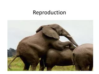
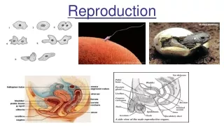
REPRODUCTION. HL. Light Micrograph of Testis. Developing spermatozoa. Leydig cells . Sertoli cells. Germinal epithelium cells. 11.4.1 Micrograph of testis. Seminiferous tubule. Structure Of Seminiferous Tubule.

E N D
REPRODUCTION HL
Light Micrograph of Testis Developing spermatozoa Leydig cells Sertoli cells Germinal epithelium cells
11.4.1 Micrograph of testis. Seminiferous tubule
Structure Of Seminiferous Tubule • Each testis is composed of a tubular structure. It is from these seminiferous tubules that sperm are produced. • From puberty these tubules will produce sperm cells throughout the life of the man. • Note the basement membrane which surrounds each tubule. • Inside the basement membrane can be seen various cells which are the stages of the developing spermatozoa. • Between the seminiferous tubules are groups of cells called interstitial or Leydig cells that produce the male sex hormone, testosterone.
Spermatogenesis • Spermatogenesis begins in the testes at puberty and continues throughout life. • Each testis consists of seminiferous tubules. These are lined by germinal epithelial cells which divide repeatedly. • Between the individual seminiferous tubules is connective tissue containing blood capillaries, together with groups of interstitial cells. These cells secrete testosterone. • The germinal epithelium cells are attached to the basement membrane, along with the nutritive cells. • Sertoli cells provide nutrition to the developing spermatozoa.
Functions • Leydig Cells: These are also known as interstitial cells and are scattered in the spaces between the seminiferous tubules. They produce tetosterone. • Sertoli cells nourish the developing sperm with nutrients. They support, nourish, and regulate the spermotogenic cells.
Wall of the Seminiferous Tubule a) Basement membrane b) Germinal epithelium (2n) which divide by mitosis to produceSpermatogonium (2n) which grow and enlarge d) Primary spermatocytes (4n) go through Meioses I to Secondary Spermatocytes (2n). Secondary Spermatocytes go through Meiosis II to produce spermatids (n) e) Sertoli cells nourish and allow the spermatids to differentiate to spermatozoa. These are released into the lumen
Spermatogenesis • http://highered.mcgraw-hill.com/sites/0072495855/student_view0/chapter28/animation__spermatogenesis__quiz_1_.html
Stages involved in spermatogenesis • Multiplication phase: During this stage the spermatogonial cells divide by mitoisto produce many cells with the potential to become gametes. • Second stage is the growth phase during which the developing sex cell undergoes growth . • The third phase is the maturation phase or meiotic phase: The primary spermatocyte undergoes the first meiotic divison to give rise to haplidseconaryspermatocyte. And then the secondary spermatocyte undergoes the second meiotic division to form four spermatids
Spermiogenesis • Spermatids undergo differentiation to form to form spermatozoa. They form the cellular structures characterisitc of a spermatozoan. • These structures include flagellum for motility, an acrosome to contain enzymes necessary for fertilization.
Role of hormones in Spermatogenesis • Three hormones play role in spermatogenesis: • Follicle Stimulating Hormone (FSH) • Luteinizing Hormone (LH) • Testosterone • FSH and LH are produced in the pituitary gland and Testosterone is produced by the Leydig cells in the testis.
FSH • Stimulates sperm production in seminiferous tubules • Stimulates division and maturation of sertoli cells LH Stimulates the interstitial cells (Leydig cells) to produce testosterone. Testosterone Promotes spermiogenesis (maturation of spermatids into spermatozoa)
Structure of the human sperm The human sperm has a head with a haploid nucleus. Atop the nucleus lies a specialized lysosome called the acrosome. It contains hydrolytic enzymes that will dissolve protective layers around the egg and enable the sperm to enter and fertilize it. Behind the head is the midpiece, which is packed with mitochondria. These organelles provide the energy needed to move the tail. Whiplikemiovement of the tail, which is really a long flagellum, propel the sperm through the female reproductive tract. The small structure allows the Sperm to swim great distances.
-- • The unequal division of the cytoplasm during meiosis (oogenesis ensures that one cell only would receive virtually all of the cytoplasm, nutrients, and organelles necessary to start a new life. The cytoplasm of the ovum also contains cortical granules which function immediately after fertilization.