Download
1 / 16
350 likes | 1.46k Vues
Viral Hemorrhagic fever: . The viral hemorrhagic fevers ( VHFs ) are a diverse group of animal and human illnesses that are caused by four different families of RNA viruses: 1- Arenaviridae . 2- Filoviridae . 3- Bunyaviridae . 4- Flaviviridae .

E N D
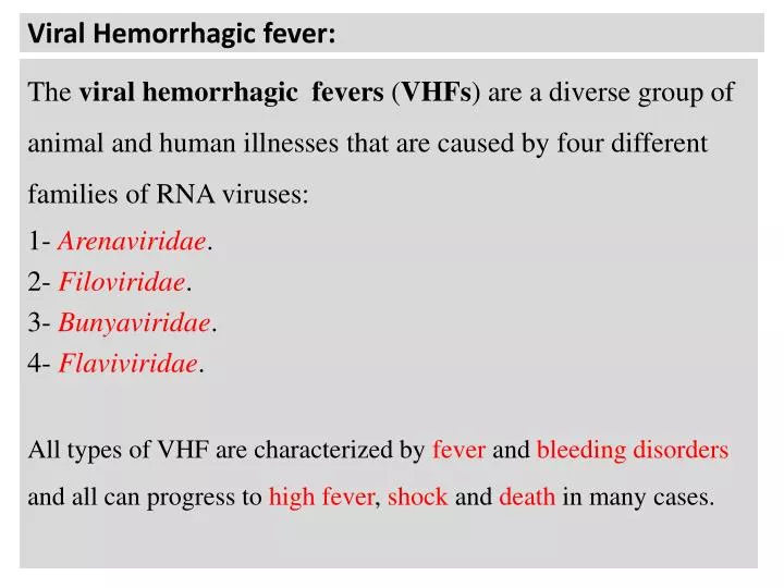
Viral Hemorrhagic fever: The viral hemorrhagicfevers (VHFs) are a diverse group of animal and human illnesses that are caused by four different families of RNA viruses: 1- Arenaviridae. 2- Filoviridae. 3- Bunyaviridae. 4- Flaviviridae. All types of VHF are characterized by fever and bleeding disorders and all can progress to high fever, shock and death in many cases.
Arenaviridae: Lassavirus, Juninvirus, and Machupo virus (MACV) are associated with hemorrhagic fever. Epidemiology: Lassa virus: West Africa. Junin virus: Argentina. Machupo virus: Bolivia. Virology: -These are round, pleomorphic, and enveloped with a diameter of 120 nm. - Nucleocapsid with two single-stranded RNA circular segments.
N Viral reservoir: Some Arenavirusesare zoonotic pathogens and are generally associated with rodentstransmitted disease. Transmission: Humans could be infected through mucosal exposure to aerosols, or by direct contact of broken skin with the infectious material, derived from infected rodents. Ingestion of contaminated food, person-to-person contact is incriminated with transmission of the virus in some cases.
n Pathogenesis and clinical picture: Incubation period: 10-14 days. The viruses infect macrophages, carried to bloodvessels ; endothelialcells infection, T-cell mediated response, vascular damage. Visceral hemorrhage. Liver and spleen necrosis. hemorrhage shock and cardiac damage. Deathin 50% of cases.
Filoviridae: Marburg virus and Ebola virus : Epidemiology : Endemic in Africa. 1-Marburgvirus (Zimbabwe and Kenya). 2-Ebolavirus (Sudan and Zaire). Reservoir: African green Monkeysand Wild Monkeys. Transmission: Monkeys-Human contact, human-human contact, and unknown routes of transmission. -The first infection was first detected among Laboratory workersin Marburg, who had been exposed to tissue culture of cell line prepared from Monkeys.
Virology: Filoviruses have a Single stranded RNA, which encodes seven proteins Envelopedfilamentous virus with Helicalnucleocapsid measuring 800 to as long as 14000nm.
Pathogenesis and clinical picture: -Eosinophiliccytoplasmicinclusions are seen in cells infected with the virus. -These virus infection disseminated with blood stream to parenchymalcells of liver, spleen, lymphnodes, and lung. -Widespread hemorrhage causes edema and hypovolemic shock. -Liverfunctiontests: SGPT, and alkaline phosphatase elevated. - Spleen destruction and lymph node enlargements.
Bunyaviridae: -Super-group of at least 200 different viruses. -Most are Arboviruses. Virology: Enveloped, spherical virion with a diameter of 90-100 nm. Single-stranded RNA nucleocapsids virus.
N Transmission: Vector-born infection 1-Phlebovirus: Sandfly, genus : Phlebotomus. 2-Nairovirus: Ticks. 3-Bunyavirus: Mosquito. Vertebrate host: 1-Phlebovirus: Sheep, cattle, others 2-Nairovirus: cattle, goats 3-Bunyavirus: Rodents, small mammals, and birds. Disease: 1-Rift Valley fever ; hemorrhagic fever, encephalitis. 2-Crimean-Congo -Hemorrhagic fever. 3-California encephalitis.
Pathogenesis and clinical picture: -Primaryviremia; flu-like symptoms, infection of vascular endotheliumand Macrophageof Reticuloendothelial system ; secondary viremia. 1-Vascular endothelial damage; leakage of plasma and erythrocytes, hemorrhagic fever. 2- Cerebral edema and encephalitis. 3- Kidney hemorrhagic necrosis.
Flaviviridae: Flavivirusesare Arboviruses transmitted to man by vector (flavus (Latin term)means yellow). Two types of Flaviviridaecause Hemorrhagicfever (H.F), and Yellowfever: 1-Dengue virus: DHF: dengue H.F, and DSS: dengue shock syndrome. 2-Yellow fever virus: Yellow fever and Hepatitis. Vector: Mosquito of the genus Aedes. Host: Human and Monkeys.(Zoonosis). Distribution: Dengue fever: Worldwide, especially tropics. Yellow fever: Africa, and South America.
Virology: - Positive Single-strandedRNA viruses. - Enveloped, Icosahedralnucleocapsid. - Size: 40-65 nm. Glycoprotein spikes (adhesion of virus to tissue). -All Flavivirusesare serologically related: SoAnti-Virus serotype antibodies could be used as prophylactic vaccine for others.
Pathogenesis and clinical picture: - Biting of the host skin by vector ( FemaleMosquito). , inoculation of smallcapillariesbloodstream by virus in Saliva. -The virus will be carried by dendriticcells throughout small capillaries to the targettissue. -Target tissue: 1-The endothelial cells of Capillaries. 2-The blood Macrophage and monocytes. -Blood phagocytes will transfer the microbe to the Reticuloendothelialsystem(RES).
N This will initiate Primary Viremia (fever, chill, headaches, and flu-like symptoms within 3-7 days). Secondary Viremia is associated with efficient replication of virus in RES. -This viremia can produce sufficient virus to infect: 1-Liver: Hepatitis, and Jaundice; yellow fever. 2-Brain: Encephalitis. 3-Vasculature and skin: Hemorrhage and shock.