Download
1 / 64
660 likes | 916 Vues
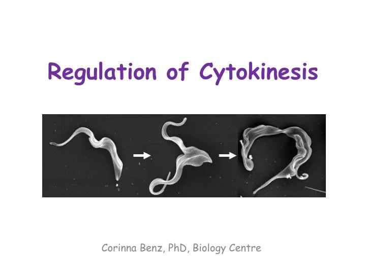
Regulation of Cytokinesis. Corinna Benz, PhD, Biology Centre. Overview. Cell cycle and cytokinesis in mammalian cells Cell cycle and cytokinesis in trypanosomes NDR kinase – MOB signalling in different organisms. General idea. The eukaryotic cell cycle.

E N D
Regulation of Cytokinesis Corinna Benz, PhD, Biology Centre
Overview • Cell cycle and cytokinesis in mammalian cells • Cell cycle and cytokinesis in trypanosomes • NDR kinase – MOB signalling in different organisms
The eukaryotic cell cycle Alberts et al., Molecular Biology of the Cell, 4th edition
Different microtubules in a mammalian spindle Alberts et al., Molecular Biology of the Cell, 4th edition
A condensed chromosome with attached kinetochore microtubules Alberts et al., Molecular Biology of the Cell, 4th edition
Cell cycle control system • Events must occur in a timely fashion • Correct order • Only once per cell cycle • Backup system if things go wrong (checkpoints) • Adaptability
Cell cycle checkpoints Alberts et al., Molecular Biology of the Cell, 4th edition
The major players – Cyclin-dependent protein kinases (Cdks) • Oscillation between active and inactive states cyclical phosphorylation of cellular proteins that initiate or regulate cell cycle events • Activity regulated by binding to a cyclin partner (with exceptions) • Cyclins are degraded cyclically while Cdks are constitutively present
Cyclin-dependent protein kinases (Cdks) and cyclins M-Cdk promotes mitotic events G1-Cdk promotes passage through a restriction point G1/S-Cdk commits cell to DNA replication S-Cdk is required for initiation of DNA replication Alberts et al., Molecular Biology of the Cell, 4th edition
The major cyclins and Cdks of vertebrates and budding yeast Vertebrates Budding Yeast • * There are three D cyclins in mammals (cyclins D1, D2, and D3). • ** The original name of Cdk1 was Cdc2 in both vertebrates and fission yeast, • and Cdc28 in budding yeast Adapted from Alberts et al., Molecular Biology of the Cell, 4th edition
Regulation via phosphorylation • Cdk-cyclin complexes are regulated through phosphorylation • Particularly important at onset of mitosis • Further regulation through Cdk inhibitor proteins (CKIs) Alberts et al., Molecular Biology of the Cell, 4th edition
Control through proteolysis • SCF (Skp1-Cullin-F-box protein): Ubiquitinligase, active in G1/S, constitutively active, substrates become available through phosphorylation • APC/C (anaphase-promoting complex): Ubiquitinligase, active in G1 (subunit Cdh1) and M (subunit Cdc20), activated by addition of subunits APC/C complex SCF complex
Control through proteolysis Alberts et al., Molecular Biology of the Cell, 4th edition
Exit from G0 and G1 events Cdk2:cyclin E G1/S-Cdk Cdk7 P Rb E2f + G1 S growth factor stimulus cyclin D ↑ G0 to G1 transition Cdk4:cyclin D/Cdk6:cyclin D G1-Cdk Rb- negative feedback loop cyclin A/B transcription of e.g. cyclin A cyclin B cyclin E E2f cyclin E ↑ APC/CCdh1 cyclin A/B-ubiquitin degradation
Exit from G0 and G1 events • Most cells in adults are in quiescent G0 stage • Growth factor stimulus cyclin D levels increase and Cdk4:cyclin D/Cdk6:cyclin D (G1-Cdk) drives cells into G1 by phosphorylating retinoblastoma tumor suppressor protein (Rb) • PhosphorylatedRb can no longer inhibit transcription factor E2f expression of genes important for DNA replication is induced • These genes include cyclin A, B and E: Only cyclin E accumulates in G1 since cyclin A and B are marked for degradation by APC/CCdh1 • Cdk2:cyclin E (G1/S-Cdk) promotes G1-S transition
Genome duplication: Replication licensing 1) Helicase loader Mcmhelicase (inactive) DePamphilis et al, Frontiers in PHYSIOLOGY, 2012
Genome duplication • Initiated at replication origin, where a preRC (pre-replication complex) is assembled which is then converted to a preIC (pre-initiation complex) • Replication licensing = Assembly of preRCs on chromatin • Replisome: DNA replication proteins • preRC assembly occurs only when Cdk activity is low (anaphase to G1/S transition) • 1) Assembly of helicase loader: Orc1-6, Cdc6 and Cdt1 • 2) Assembly of Mcmhelicase (inactive): Mcm2-7 (double hexamer)
Replication initiation 1) 2) DePamphilis et al, Frontiers in PHYSIOLOGY, 2012
Replication initiation 3) 4) DePamphilis et al, Frontiers in PHYSIOLOGY, 2012
Replication initiation 5) DePamphilis et al, Frontiers in PHYSIOLOGY, 2012
Replication initiation • DDK (Dbf4-dependent Cdc7 kinase) phosphorylates Mcm4 and 6 • Sld3 and Cdc45 assemble into preRC • Sld2 and 3 are phosphorylated by Cdk2:cyclin E (G1/S-Cdk) • Sld2-P and Sld3-P bind to Dpb11 recruitment of GINS complex • Recruitment of Pol-ε (leading strand synthesis) • ssDNA binding protein RPA required to facilitate DNA unwinding • Formation of two active replisomes upon addition of Pol-α:primase and Pol-δ • Mcm10 stabilises final replisome structure
DNA replication checkpoints 2) 3) DePamphilis et al, Frontiers in PHYSIOLOGY, 2012
DNA replication checkpoints • Effectorkinase: Chk1 • Inhibits activation of preRCs by: • Phosphorylating and inhibiting phosphatase Cdc25, which then prevents activation of Cdk2 • Phosphorylating Sld3 (at a site different from the one phosphorylated by Cdk2:cyclin E (G1/S-Cdk)), which prevents interaction with Dpb11 (prevents formation of Cdc45:Mcm:GINS complex)
Prevention of DNA re-replication DePamphilis et al, Frontiers in PHYSIOLOGY, 2012
Prevention of DNA re-replication • Cdk2:cyclin A (S-Cdk) phosphorylates Orc1, Cdc6 and Cdt1 export to cytoplasm and/or degradation • Absence of Orc1 causes other Orc subunits to leave the complex • Non-phosphorylated Cdt1 bound to PCNA (polymerase processivity factor) is ubiquitinated by CRL4:Cdt2 • Re-expression of preRC proteins in G2 and M phase, but in modified forms which are kept inactive • Metaphase exit: Cdc14 de-phosphorylatesOrc subunits and Cdc6 allowing preRC assembly • Geminin degraded Cdt1 becomes available for preRC assembly
DNA damage response Wang et al., Molecular Cancer, 2009
DNA damage response • Checkpoint kinases Chk1 and Chk2, activated by ATR and ATM kinases • ATM activated by ionising radiation • Chk2 in ATM-Chk2-Cdc25 pathway that senses double strand breaks, not essential in mammals • ATR activated by UV radiation • Chk1 in Atr-Chk1-Cdc25 pathway that senses single strand breaks, bulky lesions and stalled replication forks, essential in mammals • Both pathways result in Cdc25 phosphorylation sequestered in cytoplasm by 14-3-3 M-Cdk inactive • p53 phosphorylation causes activation of p21 (M-Cdk inhibitor) and upregulation of 14-3-3
Exit from S phase P P P CRL1:Skp2 cyclin E-P cyclin E-P -ubiquitin DNA replication proteins ↓ preRC proteins ↓ Cdk2:cyclin A S-Cdk E2f-P translocates to nucleus phosphorylates substrates to initiate mitosis Cdk1:cyclin B M-Cdk Cdk1:cyclin B-P
Exit from S phase • Cyclin E inactivated through phosphorylation by Cdk2:cyclin A (S-Cdk) becomes a target for CRL1:Skp2 ubiquitinligase and is degraded • Cdk2:cyclin A (S-Cdk) also phosphorylates and inactivates E2f (downregulation of expression of genes required for DNA replication, also prevents re-licensing of origins) • Cyclin B levels increase Cdk1:cyclin B (M-Cdk) complex formed, translocates to nucleus to phosphorylate substrates important for entry into mitosis
Entry into mitosis – M-Cdk activation P P Wee1 Myt1 Y15-P Y15-P Cdc25 Cdk1 Cdk1 Cdk1:cyclin B M-Cdk Positive feedback loop T14-P T14-P G1, S, G2 M G1
Entry into mitosis – APC/C activation P P S/G2: APC subunits: * Cdc20-P active * Cdh1-P inactive Emi1 Cdc20-P Cdk2:cyclin A S-Cdk APC/C Cdh1 binding degradation Emi1 Prometaphase: Cdk1:cyclin B M-Cdk Cdc20-P APC/CCdc20 cyclin A Cdk2 Entry into mitosis APC/CCdc20 p21/p27 cyclin B Cdk1 Anaphase progression
Entry into mitosis • Positive feedback loop increases activity of Cdk1:cyclinB (M-Cdk) • APC/C regulation: • Cdc20 subunit is active when phosphorylated • Cdh1 subunit is inactive when phosphorylated • During S and G2: APC/C inactive, Emi1 prevents phosphorylation of Cdc20 and Cdk2:cyclin A (S-Cdk) phosphorylates the APC/C, which inhibits binding of Cdh1 and results in its degradation • Prometaphase: Selective Emi1 degradation • Negative feedback loops regulate Cdk1/2 activity through APC/C-mediated ubiqitination of cyclin A and B • Cdk1 and Cdk2 also regulated by interaction with Cdk inhibitors (CKIs) p21 and p17 whose expression follows that of the APC/C
M-Cdk – functions • Induces assembly of mitotic spindle • Ensures that replicated chromosomes attach to spindle • In many organisms also triggers chromosome condensation, nuclear envelope breakdown, actin cytoskeleton rearrangement, reorganisation of Golgi and ER; e.g. through phosphorylation of lamins dismantling of nuclear envelope
Metaphase-anaphase transition Alberts et al., Molecular Biology of the Cell, 4th edition
Metaphase-anaphase transition • M-Cdkphosphorylatescondensin complex resulting in chromosome condensation at prometaphase • M-Cdk activates APC/CCdc20 triggers anaphase by promoting degradation of securin • Securin no longer inhibits the protease separase • Separase becomes active and cleaves cohesin subunits resulting in sister chromatid separation
Spindle attachment checkpoint • Mitotic spindle in green • Mad2 in red • Sister chromatids only separated when all chromosomes properly attached to spindle • State of kinetochore monitored • Several proteins e.g. Mad2 recruited to unattached kinetochores inhibition of APC/CCdc20 Alberts et al., Molecular Biology of the Cell, 4th edition
Cytokinesis Agromayor and Martin-Serrano, TRENDS in Cell Biology, 2013
Cytokinesis initiation • Signalling between anaphase spindle and cortex • Spindle recruits narrow zone of active RhoA (GTPase) • Active RhoA recruits effector contractile ring proteins (cytokinesisformin, Rhokinase, Citron kinase) • RhoA flux model: Global GAP-mediated RhoA inhibition versus localised GEF-mediated RhoA activation (e.g. Ect2 at cell equator) Green et al, Annu. Rev. Cell Dev. Biol., 2012 GAP=GTPase activating protein, GEF=Guanine nucleotide exchange factor
Central spindle (spindle midzone) formation • Spindle midzone: Overlapping, antiparallel microtubules (MTs) (+ ends facing each other) • Formation requires PRC1 and kinesins Kif4 and MKLP1 • PRC1: antiparallel MT cross linker • Chromosomal Passenger Complex (CPC): Aurora B (kinase) and three additional subunits phosphorylates and recruits • Centralspindlin: Heterotetramer, consists of two molecules MKLP1 and two molecules CYK-4 (GAP) Green et al, Annu. Rev. Cell Dev. Biol., 2012
Central spindle (spindle midzone) formation • PRC1 recruits Kif4 to overlap zones where it slows down MT dynamics • Polo-like kinase 1 (Plk1) required for spindle elongation • Plk1 recruits itself by phosphorylating substrates (e.g. PRC1, MKLP2) and thus creating binding sites • Plk1 phosphorylates CYK-4 subunit of centralspindlin, which then scaffolds recruitment of Ect2 Green et al, Annu. Rev. Cell Dev. Biol., 2012
Central spindle (spindle midzone) formation • Ect2 (GEF for RhoA) converts RhoA-GDP into RhoA-GTP, which triggers contractile ring assembly • Contractile ring consists of actin, myosin II and septin filaments (recruited by anillin which as a crosslinker binds to all three) • Ring is disassembled as it constricts Green et al, Annu. Rev. Cell Dev. Biol., 2012
Spindle midzone midbody • Upon contractile ring constriction, midzone-localised proteins are redistributed: • PRC1 and Kif4 stay at MT overlap zone • Centralspindlin and Ect2 concentrate in midbody ring, where they colocalise with anillin, RhoA, ARF6 and Cep55 • CENP-E, MKLP2 and Aurora B colocalise with tightly packed, parallel midbodyMTs in regions flanking the midbody core • Plk1 essential for these relocalisations Green et al, Annu. Rev. Cell Dev. Biol., 2012
Contractile ring midbody ring • Anillin required for assembly of midbody ring and anchoring to plasma membrane • Citron kinase essential for abscission, required for localisation of anillin and RhoA • RhoA required for anillin localisation to midbody ring Green et al, Annu. Rev. Cell Dev. Biol., 2012
Abscission Agromayor and Martin-Serrano, TRENDS in Cell Biology, 2013
Abscission • CEP55, TSG101 and ALIX are translocated to midbody and mediate recruitment and polymerisation of ESCRT-III (complex for scission) • MIT-domain containing protein 1 (MITD1) coordinates activity of ESCRT-III • Rab35 recruits OCRL and Rab11/FIP3-positive endosomes deliver p50RhoGAP to midbody resulting in changes of membrane lipid composition and clearing of actin • FYVE Cent interacts with PtdInsP and recruits TTC19 and CHMP4B • ESCRT-III filaments constrict midbody • Spastin (MT severing enyzme) cleaves microtubules • AAA ATPase VPS4 disassembles ESCRT-III
Abscission checkpoint Agromayor and Martin-Serrano, TRENDS in Cell Biology, 2013
Abscission checkpoint • CHMP4C (ESCRT-III subunit) regulates Aurora B-mediated abscission checkpoint • Trapped chromatin at midbody CHMP4C binds to Borealin and is phosphorylated by Aurora B • CHMP4C-P relocalises to midbody preventing abscission until chromatin is removed