Download
1 / 32
320 likes | 821 Vues
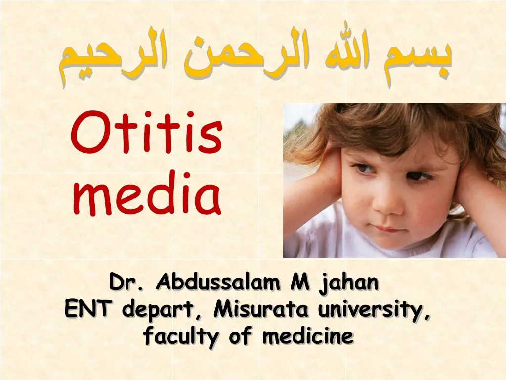
بسم الله الرحمن الرحيم. Otitis media. Dr. Abdussalam M jahan ENT depart, Misurata university, faculty of medicine. Anatomy of the Ear. Inflammation of Middle ear. Acute OM. A non supp OM. A supp. OM. A necrotizing OM. Chronic OM. Chronic supp. OM. Chronic non Supp.OM. Acute OM.

E N D
بسم الله الرحمن الرحيم Otitis media Dr. Abdussalam M jahan ENT depart, Misurata university, faculty of medicine
Inflammation of Middle ear Acute OM A non supp OM A supp. OM A necrotizing OM Chronic OM Chronic supp. OM Chronic non Supp.OM
Acute OM Acute inflammation of mucoperiosteal lining of middle ear cleft. It is common disease especially in children.. Why. (ET, URTI, AdT, bottle feeding) .
Bacteriology: Streptococcus Hemolyticus Streptococcus pnumoniae Homophiles influenza Route ET Perforated TM
Stages of AOM Tubal occlusion ET obstruction. → –ve pr. → ME mucosa swollen and hyperaemic. Catarrhal stage ME full of secretion Suppuration stage rupture of TM → pus come out Stage of resolution ME retained Normal
C/P of AOM Tubal occlusion Fullness of ear + mild otalgia Catarrhal stage increase pain + fever Suppuration stage sever pain, high fever, deafness discharge, pain disappear, decrease fever
Signs of AOM • Congested TM. • Pulging of TM. • Deafness. • Purulent discharge. • Perforated TM.
Treatment of AOM • Ear cleaning. • Systemic AB. • Local Ear drops. • analgesia. • Myringotomy +/-.
Chronic OM Chronic supp. OM Chronic non Supp.OM Safe ( Tubotympanic) Unsafe (Attico antral)
CSOM Chronic inflammation of middle ear cleft (ME cavity, ET, Mastoid). It is common disease in low socioeconomic classes.
Safe ( Tubotympanic) Unsafe (Attico antral) Definition: it is an epithelial cyst that contains keratin presented in middle ear ( presence of skin in a wrong place) . cholesteatoma
Tubotympanic(safe) Microbiology: Grame +ve bacteria Chronic inflammation Involve the ant part of ME ( tympanic cavity + ET)
S/S of CSOM safe • otorrhea. • -profuse • - mucopurulent. • - odorless. • - on / off. • Deafness ( -ve Rinne test). • TM perforation.. central.
Treatment of safe CSOM • Cleaning ( suction or mopping). • Systemic AB. • Local ear drops. • Surgical intervention: • myringoplasty
Attico antral(unsafe) Microbiology: Grame -ve bacteria & anaerobic Chronic inflammation Involve the post part of ME ( attic, antrum, mastoid)
Attico antral(unsafe) Cholesteatoma Definition: it is an epithelial cyst that contains keratin presented in middle ear ( presence of skin in a wrong place) .
Cholesteatoma Acquired Congenital primary secondary
S/S of CSOM unsafe • otorrhea. • -scanty. • - purulent. • - offensive. • - continuous. • Deafness ( -ve Rinne test). • TM perforation, marginal. • Polypi or granulation tissues.
Treatment of unsafe CSOM • Only Surgical intervention: • Mastoidectomy • - Ct scan is important in management of CSOM with Cholesteatoma.
Difference between safe and unsafe OM cholesteatoma Yes No discharge Profuse scanty, offensive perforation central peripheral Site of infection tympanic cavity, ET attic , Antrum Treatment medical or surgical always surgical
Complications of CSOM Cranial Intracranial Extracranial
Chronic non Supp.OM Secretory OM or OM with effusion ( glue ear) definition: Collection of fluids behind intact TM with out s/s of inflammation. Common in children under 9 yr. etiology: - ET dysfunction. - post unresolved AOM.
ET dysfunction Secretional Mechanical Functional Increase compliance as in children Adenoid -Deficient mucociliary clearance as in Kartageners syndrome -Abnormal viscid sec: Mucoviscidosis NP tumours Tubal oedema as post RT Abnormal opening mechanism as in Cleft palate. Tubal scaring as post Ad
So causes of SOM • Adenoid hypertrophy. • NP carcinoma. • Post AOM. • Abnormal viscid sec. • Abnormal ciliary function. • Post op scaring
symptoms of SOM • Hearing loss. • Feeling of blockage. • tinnitus. • Symptoms of cause.
Signs of SOM • Retracted TM. • Fluid level ( hair line). • Air bubbles. • TFT → CHL.
Investigations of SOM • Tympanometry. • PTA. • X- ray NP. • CT scan if needed.
medical Treatment of SOM • mucolytic. • Steroids. • Decongestant N drop. • Valsalva. surgical • Ventilation tube ( Grommet). • Adenoidectomy
Chronic non Supp.OM Adhesive OM definition: It is a complication of SOM, in which the TM become thin, atrophic, and adherent to the middle ear structures. treatment: - grommet. - tympanoplasty.
clinical case: 5 yrs old child presented with: Nasal obstruction + night snoring • Chronic nasal discharge. • Decrease hearing. • On examination: • Retracted TM • Fluid behind TM • TM not perforated. • What is Dx and how to • confirm .
THANK YOU Please visit: www.Zahrawi.Ly