Download
1 / 122
1.22k likes | 1.25k Vues
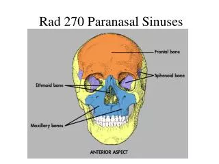
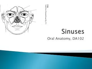
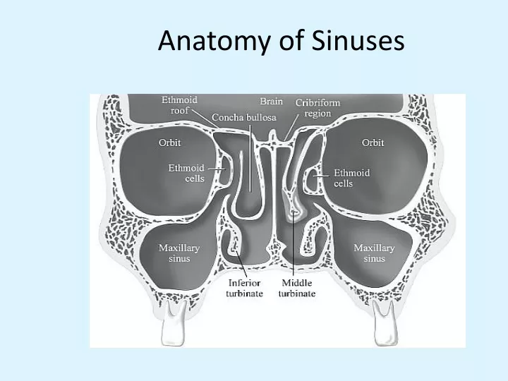
Anatomy of Sinuses. Acute Rhinosinusitis (Viral). Common Symptoms: Nasal discharge, nasal congestion, facial pressure, cough, fever, muscle aches, joint pains, sore throat with hoarseness. Symptoms resolve in 10-14 days Common in fall, winter and spring. Treatment: Symptomatic.

E N D
Acute Rhinosinusitis (Viral) Common Symptoms: Nasal discharge, nasal congestion, facial pressure, cough, fever, muscle aches, joint pains, sore throat with hoarseness. Symptoms resolve in 10-14 days Common in fall, winter and spring. Treatment: Symptomatic
Acute Bacterial Sinusitis • Causative agents are usually the normal inhabitants of the respiratory tract. • Common agents: Streptococcus pneumoniae Nontypeable Haemophilus Influenzae Moraxella Catarrhalis
Diagnosis Based on clinical signs and symptoms Physical Exam: Palpate over the sinuses, look for structural abnormalities like DNS. X-ray sinuses: not usually needed but may show cloudiness and air fluid levels Limited coronal CT are more sensitive to inflammatory changes and bone destruction
Signs and Symptoms Feeling of fullness and pressure over the involved sinuses, nasal congestion and purulent nasal discharge. Other associated symptoms: Sore throat, malaise, low grade fever, headache, toothache, cough > 1 week duration. Symptoms may last for more than 10-14 days.
Manifestations • nasal drainage and congestion • facial pain or pressure • headache. • Thick, purulent or discolored nasal discharge • is often thought to indicate bacterial sinusitis, but it also occurs early in viral infections such as the common cold • Other nonspecific symptoms include cough, sneezing, and fever • Tooth pain, most often involving the upper molars, is associated with bacterial sinusitis Dr. Farzin khorvash
Manifestations • sinus pain or pressure often localizes and be worse when the patient bends over or is supine • symptoms of advanced sphenoid or ethmoid sinus: severe frontal or retroorbital pain radiating to the occiput, thrombosis of the cavernous sinus, and signs of orbital cellulitis • advanced frontal sinusitis ,Pott's puffy tumor, swelling and pitting edema over the frontal bone ,subperiosteal abscess Dr. Farzin khorvash
Diagnosis • illness duration • acute bacterial sinusitis is uncommon in patients whose symptoms have lasted <7 days • facial or tooth pain in combination with purulent nasal discharge that have persisted for >7 days Dr. Farzin khorvash
Diagnosis and Management of Acute Sinusitis • Update of 2001 guideline • Focuses on ages 1–18 years • Not subacute or chronic; not <1 year • Not anatomic abnormalities; immunodeficiencies, cystic fibrosis, ciliary dyskinesia
Diagnosis and Management of Acute Sinusitis Areas of change: • Addition of “worsening course” • New data on effectiveness of antibiotics • Option to observe for 3 days in “persistent” infection • Imaging is not necessary to identify or confirm a diagnosis of acute sinusitis
Key Action Statement 1 Clinicians should make a diagnosis of acute bacterial sinusitis (ABS) when a child with an upper respiratory infection (URI) presents with: • Persistent illness (nasal discharge or daytime cough or both for ≥10 days without improvement) • Worsening course (worsening or new onset of nasal discharge, daytime cough or fever after initial improvement) • Severe onset (concurrent fever and purulent nasal discharge for 3 days)
Severe Worsening Common Clinical Presentations for ABS Persistent Symptoms
Acute Sinusitis “Persistent Symptoms” • 10–30 days (no improvement) • Nasal discharge (any quality) • Daytime cough (worse at night) • Fever – variable • Headache and facial pain – variable
Persistent Symptoms • Only 6–8% of children meet criteria Before concluding that child has sinusitis: • Differentiate between sequential episodes of URI and sinusitis • Establish that symptoms are NOT improving
Acute Sinusitis “Severe Symptoms” • High fever (T ≥39oC) and • Purulent nasal discharge concurrently for at least3–4 days • Need to distinguish from uncomplicated viral infections with moderate illness
“Worsening Symptoms” • Typical viral URI symptoms • Nasal discharge or cough or both for 5–6 days which is improving • Sudden worsening manifests as • Increase nasal discharge or cough or both • Onset of severe headache • Onset of new fever
Images – Key Action Statement 2A Clinicians should not obtain imaging studies (plain x-rays, computed tomography [CT] , magnetic resonance imaging [MRI] or ultrasound [U/S]) to distinguish ABS from viral URI Brian Evans/Photo Researchers/Getty Images
Images • Historically, imaging was confirmatory • No longer recommended • Continuity of respiratory mucosa leads to diffuse inflammation during viral URI • Responsible for controversy regarding images
computed tomography, sinus radiography • patients who meet these criteria, only 40 to 50% have true bacterial sinusitis • CT or XR is not recommended for routine cases, particularly early in the course of illness (i.e., at <7 days) • persistent, recurrent, or chronic sinusitis, CT of the sinuses is choice. Dr. Farzin khorvash
Imaging of Sinuses • 1940s – Observations made regarding frequency of abnormal sinus radiographs in “healthy” children • 1970s and 1980s – Children with URI had frequent abnormalities of paranasalsinuses • As CT scanning of central nervous system (CNS) and skull became prevalent, incidental abnormalities observed • When MRI performed in children with URI, 70% show major abnormalities of mucosa
Computed Tomographic Study of the Common Cold • 31 healthy young adults with new “cold” • Recruited within 48–96 hours • To have CT of paranasal sinuses • 87% had significant abnormalities of their maxillary sinuses; with air-fluid level • Conclusion: Common cold associated with frequent and striking abnormalities of sinuses Gwaltney JM Jr, Phillips CD, Miller RD, et al. Computed tomography study of the common cold. N Engl J Med. 1994;330(1):25–30
Coronal computed tomographic scan showing ethmoidal polyps. Ethmoid opacity is total as a result of nasal polyps, with a secondary fluid level in the left maxillary antrum.
Abnormalities on CT Scan Image provided by speaker.
Summary of Imaging • When paranasal sinuses are imaged in any way in children with uncomplicated URI, majority will be significantly abnormal • Normal images = No sinusitis • Abnormal images cannot confirm diagnosis and are not necessary in children with uncomplicated clinical sinusitis
Images – Key Action Statement 2B Clinicians should obtain a contrast-enhanced CT scanof the paranasal sinuses and/or an MRI with contrast whenever a child is suspected of having orbital or CNS complications of ABS
Complications of Sinusitis Orbital a. sympathetic effusion b. subperiosteal abscess c. orbital abscess d. orbital cellulitis e. cavernous sinus thrombosis
Orbital Complications of Sinusitis • Proptosis – anterior and lateral displacement of globe • Impairment of extraocular movements • Loss of visual acuity • Chemosis – edema of conjunctiva
CNS Complications of ABS Suspected with very severe headache, photophobia, seizure, other focal neurologic findings • Subdural empyema • Epidural empyema • Venous thrombosis • Brain abscess • Meningitis
Initial Management of ABS • Key Action Statement 3A: Clinician should prescribe antibiotic therapy for ABS in children with severe onset or worsening course • Key Action Statement 3B: Clinician should either prescribe antibiotic therapy OR offer additional outpatient observation for 3 days to children with persistent illness
Initial Management of ABS Guidance for clinician regarding management of children with persistent symptoms: • Antibiotic therapy – starting as soon as possible after the encounter • Additional outpatient observation– for 3 days with plan to begin antibiotics if child does not improve or worsens at any time
Initial Management of ABS • Contrasts with 2001 AAP guideline • Acknowledges that although ABS is a bacterial infection • spontaneous resolution ~ common • 10 days is a guideline; no likely harm in allowing up to 3 more days in persistent onset • Reinforces antibiotic treatment as soon as possible in severeor worseningillness
TREATMENT • Most patients ,improve without antibiotic therapy • mild to moderate symptoms of <7 days' duration • facilitating sinus drainage, such as oral and topical decongestants, nasal saline lavage • in patients with a history of chronic sinusitis or allergies — nasal glucocorticoids. Dr. Farzin khorvash
antibiotics • not improve after 7 days • more severe symptoms (regardless of duration) Dr. Farzin khorvash
antibiotics • Empirical therapy ,S. pneumoniae and H. influenzae • amoxicillin • drug-resistant S. pneumoniae • Up to 10% of patients do not respond to initial antimicrobial therapy • these patients should be considered for sinus aspiration and/or lavage • prophylactic antibiotics to prevent episodes of recurrent acute bacterial sinusitis is not recommended. Dr. Farzin khorvash
Key Action Statement 4 Clinicians should prescribe amoxicillin with or without clavulanate as first-line treatment when a decision has been made to initiate antibiotic treatment of ABS
Microbiology of Acute Sinusitis • Gleaned from microbiology of acute otitis media (AOM) • Similar pathogenesis and pathophysiology • Middle ear is a paranasal sinus Brian Evans/Photo Researchers/Getty Images
Microbiology of ABS, 1984 • Streptococcus pneumoniae 30% • Haemophilus influenzae 20% • Moraxella catarrhalis 20% • Streptococcus pyogenes 4% • Sterile 25%
Suspected Microbiology of ABS, 2013 • Streptococcus pneumoniae 15–20% • Haemophilus influenzae 45–50% • Moraxella catarrhalis 10–15% • Streptococcus pyogenes5% • Sterile 25%
Antibiotic Resistance • S pneumoniae: 10–15%; can increase up to 50% • H influenzae: 10–68% • M catarrhalis: 100% • LIMITED CURRENT DATA ON MICROBIOLOGY
Treatment • About 2/3rd of patients will improve without treatment in 2 weeks. • Antibiotics: Reserved for patients who have symptoms for more than 10 days or who experience worsening symptoms. • OTC decongestant nasal sprays should be discouraged for use more than 5 days • Supportive therapy: Humidification, analgesics, antihistaminics
Treatment • Amoxicillin – traditional first-line therapy • Amoxicillin at 45 mg/kg/day in 2 doses • If high prevalence of penicillin-resistant S pneumoniae • Amoxicillin at 90 mg/kg/day in 2 doses
Treatment • Amoxicillin ineffective against beta-lactamase producing bacteria • Choices: • drug inherently resistant to beta-lactamase • combine amoxicillin with irreversible beta-lactamase inhibitor = K clavulanate