Download
1 / 55
580 likes | 950 Vues
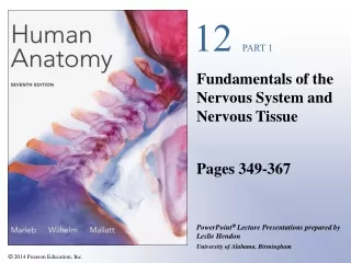
Anatomy & Physiology. The Central Nervous System. Chapter 12 Part 1. Brain Development. Posteriolateral growth of the cerebral hemispheres encloses the diencephalon and superior aspect of the brain stem. Embryo at four weeks http://w-cpc.org/fetal1.html. Embryonic Development.

E N D
Anatomy & Physiology The Central Nervous System Chapter 12 Part 1
Brain Development Posteriolateral growth of the cerebral hemispheres encloses the diencephalon and superior aspect of the brain stem
Embryonic Development Ectoderm thickens along dorsal midline to form the neural plate The neural plate invaginates, forming a neural groove flanked by neural folds The neural groove fuses dorsally and forms the neural tube which detaches from the surface ectoderm. The neural tube differentiates into the CNS. Neural crest cells form some PNS structures.
Neural Tube and Primary Brain Vesicles The anterior (rostral) end of the neural tube expands and forms the three primary brain vesicles Prosencephalon Mesencephalon Rhombencephalon
Secondary Brain Vesicles In week 5 of embryonic development, secondary brain vesicles form Telencephalon and Diencephalon arise from the prosenencephalon Mesencephalon remains undivided Metencephalon and Myelencephalon arise from the rhombencephalon
Adult Brain Structures Secondary brain vesicles differentiate into adult brain structures Telencephalon – cerebrum: cortex, white matter, and basal nuclei Diencephalon – thalamus, hypothalamus Mesencephalon – brain stem: midbrain Metencephalon – brain stem: pons Myelencephalon – brain stem: medulla oblongata
Adult Neural Canal Regions The central cavity of the neural tube enlarges in four regions to form fluid-filled Ventricles Telencephalon – Lateral Ventricles Diencephalon – Third Ventricle Mesencephalon – Cerebral Aqueduct Metencephalon & Myelencephalon – Fourth Ventricle Spinal Cord – Central Canal
Ventricles of the Brain Pair of C-shaped Lateral Ventricles Third Ventricle in the diencephalon Fourth Ventricle in the hindbrain dorsal to the pons
Ridges called Gyri (gyrus) Shallow grooves called Sulci (sulcus) Deeper grooves called Fissures Cerebral Hemispheres Form the superior part of the brain and make up over 80% of its mass Surface Features: Hemispheres are separated by the Longitudinal fissure and from cerebellum by the Transverse Cerebral fissure
Cerebral Hemispheres - Lobes Frontal, Parietal, Temporal, Occipital, and (Insula) Central sulcus – separates the frontal and parietal lobes Parieto-occipital sulcus – separates the parietal and occipital lobes Lateral sulcus – separates the parietal and temporal lobes
Cerebral Hemispheres – 3 basic regions Cortex – superficial gray matter, 1-4.5 mm thick White matter – inner, unmyelinated Basal Nuclei – islands of gray matter within the white matter Cortex (40% of brain mass) consists of neuron cell bodies, dendrites, and unmyelinated axons (plus glial cells and blood vessels), no fiber tracts, all neurons are interneurons
Cerebral Cortex Enables sensation, communication, memory, understanding, and voluntary movements Each hemisphere controls the opposite (contralateral) side of the body Hemispheres are not functionally equivalent (lateralization or specialization of cortical functions) No functional area acts alone - conscious behavior involves the entire cortex
Cerebral Cortex Three kinds of functional areas: Motor areas – control voluntary movement Sensory areas – conscious awareness of sensation Association areas – integrate diverse information, communicate “associate” with the motor cortex and sensory association areas to analyze input
Cerebral Cortex: Motor Areas Primary (somatic) motor cortex Premotor cortex Broca’s area Frontal eye field
Primary Motor Cortex Located in the precentral gyrus of the frontal lobe Allows conscious control of voluntary movements Composed of neurons called pyramidal cells - axons project to spinal cord and make up the voluntary motor tracts called corticospinal tracts
Primary Motor Cortex Motor homunculus – caricature of relative amounts of cortical tissue devoted to each motor function
Premotor Cortex Located anterior to the precentral gyrus Controls learned, repetitious, or patterned motor skills Coordinates simultaneous or sequential actions( directly or indirectly) Involved in the planning of movements
Broca’s Area Located anterior to the inferior region of the premotor area Present in one hemisphere (usually the left) A motor speech area that directs muscles involved in speech Is active as one prepares to speak
Frontal Eye Field Located anterior to the premotor cortex and superior to Broca’s area Controls voluntary eye movement
Sensory Areas Primary somatosensory cortex Somatosensory association cortex Visual and auditory areas Olfactory, gustatory, and vestibular cortices
Primary Somatosensory Cortex Located in the postcentral gyrus of the parietal lobe Receives information from somatic sensory receptors in the skin and proprioceptors in skeletal muscles Exhibits spatial discrimination
Primary Somatosensory Cortex Somatosensory homunculus – caricature of relative amounts of cortical tissue devoted to each sensory function
Somatosensory Association Cortex Located posterior to the primary somatosensory cortex Integrates sensory information coming from the primary somatosensory cortex to produce an understanding (size, texture, and relationship of parts) of the stimulus
Visual Areas Primary visual (striate) cortex - located on the posterior tip of the occipital lobe, most of it is buried in the calcarine sulcus - receives visual information from the retinas Visual association area - surrounds the primary visual cortex - interprets visual stimuli (e.g., color, form, and movement)
Auditory Areas Primary auditory cortex - located in the superior margin of the temporal lobe -receives information related to pitch, rhythm, and loudness Auditory association area - located posterior to the primary auditory cortex -stores memories of sounds and permits perception of sounds
Other Sensory Areas Olfactory (smell) cortex – small area of frontal lobe above the orbit and in medial temporal lobe (piriform lobe and uncus) Gustatory (taste) cortex – in parietal lobe deep to temporal lobe
Other Association Areas Prefrontal cortex Language areas General (common) interpretation area Visceral association area
Prefrontal Cortex Located in the anterior portion of the frontal lobe Involved with intellect, cognition, recall, personality, judgment, reasoning, and conscience Closely linked to the limbic system (emotional part of the brain)
Language Areas Wernicke’s area Broca’s area Located in a large area surrounding the left (or language-dominant) lateral sulcus Broca’s area – speech preparation and production Wernicke’s area – involved in sounding out unfamiliar words Lateral prefrontal cortex – language comprehension and word analysis. Lateral and ventral temporal lobe – coordinate auditory and visual aspects of language
General (Common) Interpretation Area (?) Ill-defined region including parts of the temporal, parietal, and occipital lobes Found in one hemisphere, usually the left Integrates incoming signals into a single thought Involved in processing spatial relationships Visceral Association Area (?) Located in the cortex of the insula Involved in conscious perception of visceral sensations
Lateralization of Cortical Function Lateralization – each hemisphere has abilities not shared with the other hemisphere Cerebral dominance – designates the hemisphere dominant for language Left hemisphere – controls language, math, and logic Right hemisphere – controls visual-spatial skills, emotion, and artistic skills
Cerebral White Matter Consists of myelinated fibers and their tracts Responsible for communication between areas of the cerebrum amd between the cerebral cortex and lower CNS centers
Cerebral White Matter Fiber Tracts Commissures – connect corresponding gray areas of the two hemispheres (corpus callosum , anterior and posterior commissures) Association fibers – connect different parts of the same hemisphere Projection fibers – enter the hemispheres from lower brain or cord centers Commissures and Association fibers – horizontal Projection fibers – vertical
Basal Nuclei Masses of gray matter found deep within the cortical white matter Corpus striatum: Caudate nucleus (amygdala- tail of caudate) Lentiform nucleus – composed of the putamen and the globus pallidus Fibers of internal capsule run between and through caudate and lentiform nuclei, giving them a striped appearance
Functions of Basal Nuclei Possible functions of basal nuclei: Influence muscular activity Regulate attention and cognition Regulate intensity of slow or stereotyped movements Inhibit antagonistic and unnecessary movement
Diencephalon Central core of the forebrain surrounded by the cerebral hemispheres Consists of three paired structures – thalamus, hypothalamus, and epithalamus Collectively, these gray matter areas enclose the third ventricle
Thalamus Paired, egg-shaped masses that form the superolateral walls of the third ventricle Connected at the midline by the interthalamic adhesion (intermediate mass) Contains four groups of nuclei - anterior, ventral, dorsal, and posterior - project and receive fibers from the cerebral cortex
Thalamus – “gateway” to the cerbral cortex Afferent impulses from all senses converge and synapse in the thalamus Impulses of similar function are sorted out, “edited”, and relayed as a group to the appropriate area of the sensory cortex or association areas All inputs ascending to the cerebral cortex pass through the thalamus Plays a key role in mediating sensation, motor activities, cortical arousal, learning, and memory