Download
1 / 29
310 likes | 545 Vues
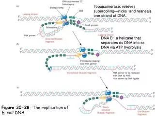
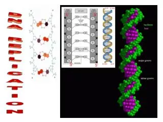
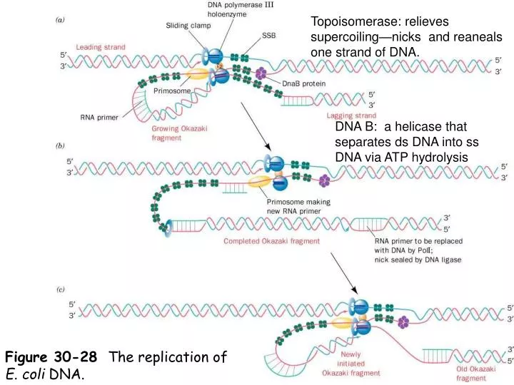
Topoisomerase: relieves supercoiling—nicks and reaneals one strand of DNA. DNA B: a helicase that separates ds DNA into ss DNA via ATP hydrolysis. Figure 30-28 The replication of E. coli DNA. Functional domains in the Klenow Fragment (left) and DNA Polymerase I (PDB).

E N D
Topoisomerase: relieves supercoiling—nicks and reaneals one strand of DNA. DNA B: a helicase that separates ds DNA into ss DNA via ATP hydrolysis Figure 30-28 The replication of E. coli DNA.
Functional domains in the Klenow Fragment (left) and DNA Polymerase I (PDB). Produced from subtilisin or trypsin cleavage Retains polymerase and 3’-5’ exo activity
Figure 30-8b X-Ray structure of E. coli DNA polymerase I Klenow fragment (KF) in complex with a dsDNA (a tube-and-arrow representation of the complex in the same orientation as Part a). Page 1141
The structure of the Klenow fragment of DNAP I from E. coli Fingers Palm
Nick Translation • Requires 5’-3’ activity of DNA pol I • Steps • At a nick (free 3’ OH) in the DNA the DNA pol I binds and digests nucleotides in a 5’-3’ direction • The DNA polymerase activity synthesizes a new DNA strand • A nick remains as the DNA pol I dissociates from the ds DNA. • The nick is closed via DNA ligase Source: Lehninger pg. 940
Figure 30-20 The reactions catalyzed by E. coli DNA ligase. Page 1150
Figure 30-21 X-Ray structure of DNA ligase from Thermus filiformis. Page 1151
Figure 30-13b The subunit of E. coli Pol III holoenzyme. Space-filling model of sliding clamp in hypothetical complex with B-DNA. Page 1146
Sliding clamp http://www.callutheran.edu/Academic_Programs/Departments/BioDev/omm/poliiib_2/poliiib.htm
Here’s a computer modelof DNA replicationhttp://www.youtube.com/watch?v=4jtmOZaIvS0 This is a pretty good outline: http://www.youtube.com/watch?v=teV62zrm2P0&NR=1 Another one with review questions (perhaps oversimplified) http://www.wiley.com/college/pratt/0471393878/student/animations/dna_replication/index.html
FIDELITY OF REPLICATION • Expect 1/103-4, get 1/108-10. • Factors • 3’5’ exonuclease activity in DNA pols • Use of “tagged” primers to initiate synthesis • Battery of repair enzymes • Cells maintain balanced levels of dNTPs
This article is a simple overview of repair processes • http://www.nature.com/nature/journal/v421/n6921/full/nature01408.html
DNA repair Ilkka Koskela Katri Vilkman
Foreword DNA • variation is an essential factor to evolution (1000-10^6 lesions per day) • stability is important for the individual (less than 1/1000 mutations are permanent) • A relatively large amount of genes are devoted to coding DNA repair functions.
Sources of damage: heat metabolic accidents (free radicals) radiation (UV, X-Ray) exposure to substances (especially aromatic compounds) Types of damage: deamination of nucleotides depurination of nucleotides oxidation of bases breaks in DNA strands
Diseases • colon cancer • cellular ultraviolet sensitivity • Werner syndrome (premature aging, retarded growth) • Bloom syndrome (sunlight hypersensitivity)
Damage of the double helix • Single strand damage • information is still backed up in the other strand • Double strand damage • no backup • can cause the chromosome to break up
Single strand repair • Base excision repair • A base-specific DNA glycosylase detects an altered base and removes it • AP endonuclease and phosphodiesterase remove sugar phosphate • DNA Polymerase fills and DNA ligase seals the nick
Single strand repair • Nucleotide excision repair • a large multienzyme compound scans the DNA strand for anomalities • upon detection a nuclease cuts the strand on both sides of the damage • DNA helicase removes the oligonucleotide • the gap is repaired by DNA polymerase and DNA ligase enzymes
Double strand repair • Nonhomologous end-joining • only in emergency situations • two broken ends of DNA are joined together • a couple of nucleotides are cut from both of the strands • ligase joins the strands together
Double strand repair • Homologous end-joining • damaged site is copied from the other chromosome by special recombination proteins
DNA repair enzymes • a lot of DNA damage -> elevated levels of repair enzymes • extreme change in cell's environment (heat, UV, radiation) activates genes that code DNA repair enzymes • For an example, heat-shock proteins are produced in heat-shock response when being subjected to high temperatures.
Cell Cycle and DNA repair • Cell cycle is delayed if there is a lot of DNA damage. • Repairing DNA as well as signals sent by damaged DNA delays progression of cell cycle. ->ensures that DNA damages are repaired before the cell divides
References • Pictures • http://www.2modern.com/index.asp?PageAction=VIEWPROD&ProdID=985 • http://www.senescence.info/WS.jpg • http://en.wikipedia.org/wiki/Dna • http://www.funpecrp.com.br/gmr/year2003/vol1-2/imagens/sim0001fig1.jpg • http://www.science.siu.edu/microbiology/micr460/PageMill%20Images/image32.gif • http://www.bio.brandeis.edu/haberlab/jehsite/images/nhejd.gif • http://www.biochemsoctrans.org/bst/029/0655/bst0290655f02.gif • http://www.antigenics.com/products/tech/hsp/images/animation.jpg • http://bioinformatics.psb.ugent.be/images/illust_cell_cycle_large.jpg