Download
1 / 58
1.32k likes | 4.22k Vues
Volume 350:1646-1654 April 15, 2004 Number 16 Management of Cirrhosis and Ascites Pere Ginès, M.D., Andrés Cárdenas, M.D., Vicente Arroyo, M.D., and Juan Rodés, M.D. Pathophysiology of Ascites

E N D
Volume 350:1646-1654April 15, 2004Number 16Management of Cirrhosis and AscitesPere Ginès, M.D., Andrés Cárdenas, M.D., Vicente Arroyo, M.D., and Juan Rodés, M.D.
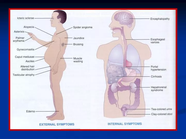
Pathophysiology of Ascites The chief factor contributing to ascites is splanchnic vasodilatation. Increased hepatic resistance to portal flow due to cirrhosis causes the gradual development of portal hypertension, collateral-vein formation, and shunting of blood to the systemic circulation. As portal hypertension develops, local production of vasodilators, mainly nitric oxide, increases, leading to splanchnic arterial vasodilatation.
In the early stages of cirrhosis, splanchnic arterial vasodilatation is moderate and has only a small effect on the effective arterial blood volume, which is maintained within normal limits through increases in plasma volume and cardiac output. In the advanced stages of cirrhosis, splanchnic arterial vasodilatation is so pronounced that the effective arterial blood volume decreases markedly, and arterial pressure falls. As a consequence, arterial pressure is maintained by homeostatic activation of vasoconstrictor and antinatriuretic factors, resulting in sodium and fluid retention.
. The combination of portal hypertension and splanchnic arterial vasodilatation alters intestinal capillary pressure and permeability, facilitating the accumulation of retained fluid within the abdominal cavity. As the disease progresses, there is marked impairment in renal excretion of free water and renal vasoconstriction — changes that lead to dilutional hyponatremia and the hepatorenal syndrome, respectively.
Figure 2. Probability of Survival among Patients with Cirrhosis, Refractory Ascites, and the Hepatorenal Syndrome. Type 1 hepatorenal syndrome is a progressive impairment in renal function, defined by a doubling of the initial serum creatinine concentration in less than two weeks to a value greater than 2.5 mg per deciliter (221 µmol per liter). Type 2 hepatorenal syndrome is a stable or slowly progressive impairment in renal function that does not meet the criterion for type 1 hepatorenal syndrome.
Spontaneous Bacterial Peritonitis Spontaneous bacterial peritonitis is characterized by the spontaneous infection of ascitic fluid in the absence of an intraabdominal source of infection. Its prevalence among patients with ascites ranges between 10 and 30 percent. The presence of at least 250 polymorphonuclear cells per cubic millimeter of ascitic fluid is diagnostic of this condition. Aerobic gram-negative bacteria, primarily Escherichia coli, are the most common isolates, although the frequency of episodes caused by gram-positive bacteria has recently increased. Spontaneous bacterial peritonitis involves the translocation of bacteria from the intestinal lumen to the lymph nodes, with subsequent bacteremia and infection of ascitic fluid. Third-generation cephalosporins are the treatment of choice.