Download
1 / 40
400 likes | 821 Vues
Can get blood from a fractious cat without actually touching it ... Smaller for kittens, puppies, toy breeds. Place the pet in dorsal recumbency and prep the ...

E N D
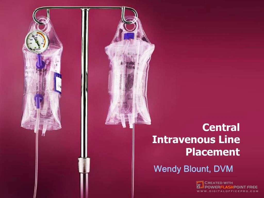
Slide 1:Central Intravenous Line Placement
Wendy Blount, DVM
Slide 2:Why Place a Central Line?
When serial blood values are needed. Avoids pain, trauma and bruising Can get blood from a fractious cat without actually touching it Makes repeated blood draws from difficult animals possible Maintains integrity of the veins for easy placement of the next IV catheter
Slide 3:Why Place a Central Line?
When do we need serial values? Diabetic ketoacidosis Renal failure requiring diuresis Liver failure hypoproteinemia �mean as a snake� diabetics who need a glucose curve Hemolytic anemia
Slide 4:Why Place a Central Line?
When you need central venous access Drugs that are caustic Doxycycline IV Total Parenteral Nutrition (TPN) Partial Parenteral Nutriiton (PPN) Monitoring Central Venous Pressure (CVP) CVP = pressure in the RA When giving IV fluids to dogs with right congestive heart failure
Slide 5:Why Place a Central Line?
Central Venous Pressure Normal 5-8 cm H20 in dogs 2-3 cm H20 in cats Increased CVP can result in signs of right heart failure >10 cm in dogs >8 cm in cats Trends are probably more important than absolute values
Slide 6:Peel Away Introducer
video
Slide 7:Guidewire Technique
video
Slide 8:Around the Needle Catheter
Standard teflon large bore IV catheter � 14-18 gauge x 3-4 inches Smaller for kittens, puppies, toy breeds Place the pet in dorsal recumbency and prep the jugular furrows If you wish, make a stab incision through skin, just medial or lateral to one of the jugular veins
Slide 9:Around the Needle Catheter
Slide 10:Around the Needle Catheter
This helps if dehydrated or hypovolemic Pass the stylet with catheter into the jugular vein Attach syringe to minimize blood spillage
Slide 11:Around the Needle Catheter
Advance catheter into the jugular vein
Slide 12:Around the Needle Catheter
Slide 13:Around the Needle Catheter
Attach extension set, so you can disconnect from IV fluids without disturbing the bandage.
Slide 14:Around the Needle Catheter
Tape or suture catheter to the skin Recover from sedation Bandage Antibiotic dressing Cast padding Roll gauze vetrap
Slide 15:Through the Needle Catheter
Angiocath � 16G or 18G Intracath � 16G or 18G Venocath � 17G or 19G
Slide 16:Through the Needle Catheter
Three layers: Guide wire stylet (inner most) Polypropylene catheter Needle stylet (outer most) Needle Guard Clamps around the needle like a clamshell, to keep the needle from cutting the catheter in half
Slide 17:Through the Needle Catheter
Occlude jugular vein at thoracic inlet Insert the needle stylet into the vein With the catheter and bag attached (below) Or with the catheter and bag detached
Slide 18:Through the Needle Catheter
When you are in the vein: See �flash� up the catheter if it is attached Blood out the needle stylet if not attached
Slide 19:Through the Needle Catheter
Stop occluding the jugular vein Decreases flow of blood out the hub if bag not attached Thread the catheter by �milking it� through the sterile bag, or threading it into the open needle hub Thread in at least 3-4 inches in a small dog or cat All the way in if dog is big enough To the right atrium, or least well into the thoracic inlet
Slide 20:Through the Needle Catheter
Place a 4x4 gauze at the venipuncture site an apply gentle pressure Withdraw the needle, leaving the catheter in place. Remove the protective bag Seat the catheter hub firmly into the needle stylet hub I like to use a drop of tissue glue to secure them together If there is movement here, the catheter can be sheared off the hub
Slide 21:Through the Needle Catheter
Apply the needle guard Secure closed with white tape Remove the wire stylet MAKE SURE the cath & needle hubs are attached (b attached to c) to each other before the stylet (a) is removed If not, you can remove the catheter (b) with the stylet (a) by mistake
Slide 22:Through the Needle Catheter
attach a 10-12cc syringe filled with saline or LRS Flush to make sure the catheter is patent Aspirate to make sure catheter is patent flush and aspirate about every minute, to make sure catheter is still patent and not kinked, while wrapping
Slide 23:Securing the Catheter
The external part of the catheter should be placed just behind the ear
Slide 24:Securing the Catheter
Place a white tape butterfly on the needle guard Secures the needle guard closed Used to suture the guard to the skin I don�t use the suture holes in the needle guard Vetafil 2-0 or other Non- absorbable suture
Slide 25:Securing the Catheter
Slide 26:Securing the Catheter
Place a small square of gauze with antibiotic ointment over the venipuncture site Change this every 2-3 days
Slide 27:Securing the Catheter
Place �-1/2 inch padding between the needle guard and the skin Cotton, gauze or cast padding - prevents sores Be careful not to kink the catheter here
Slide 28:Securing the Catheter
free flow kink
Slide 29:Securing the Catheter
REMEMBER, you are flushing and aspirating every minute or so as you go, with syringe full of saline attached to the catheter hub Secure catheter to the neck with full circle white tape Clip a �bridal path� in the fur if needed
Slide 30:Securing the Catheter
Attach the catheter hub to the outer layer of roll gauze with �split� white tape
Slide 31:Securing the Catheter
Outer layer of vetrap Cut hole over catheter Can completely cover catheter with another loop of vetrap when not in use.
Slide 32:Securing the Catheter
Attach the catheter hub to the outer layer of vetrap with �split� white tape
Slide 33:Securing the Catheter
These photos show bandaging while anesthetized. If sedation is required, I prefer to tape while sedated, then finish the bandage after awake and sitting sternal This prevents a bandage that is too tight
Slide 34:Taking Blood Samples
Disconnect IV fluid line and cap Flush the catheter with heparinized 3-5 cc saline Gently withdraw 5-6 cc of blood Gently withdraw needed sample All values except platelets will be accurate Gently replace 5-6cc of blood in �dump syringe� Flush the catheter with 3-5cc saline
Slide 35:CVP Measurement
Equipment Needed Bag of fluids Fluid administration set IV extension set Three-way stopcock Manometer If you don�t have a manometer, you cant tape IV tubing to a ruler with cm marks on it Flush patient�s IV catheter Fill the IV extension set with fluid Connect the IV extension set To patient jugular catheter at one end To 3-way stopcock at the other end (side) Stopcock off to patient
Slide 36:CVP Measurement
Slide 37:CVP Measurement
Connect manometer to 3-way stop-cock (top) This connection is the weakest point Support the manometer when turning the stopcock Connect the IV set and fill the line with fluids One end to the fluid bag (one side) The other end to the stopcock (other side)
Slide 38:CVP Measurement
Turn the stopcock off to manometer, and make sure fluid flows freely into the patient Turn stopcock off to patient, and fill manometer with fluid to at least 15-20 cm Make sure no air bubbles, which could cause vapor lock
Slide 39:CVP Measurement
Patient should be in sternal or lateral recumbency Place the -0- on the manometer at the level of the right atrium Midway between dorsal and ventral If in lateral recumbency
Slide 40:CVP Measurement
Turn stopcock off to fluids, and allow fluid to fall until it rests at the patient�s CVP The meniscus will oscillate up and down as the heart beats and the patient breathes