Download
1 / 105
1.05k likes | 1.52k Vues
Ocular disorders identified as a group due to the increase in intraocular ... Location of one ear on each side of head produces binaural hearing ...

E N D
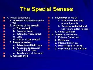
Slide 1:The Special Senses (Eye and Ear)
CHAPTER 14
Slide 2:Overview of the Eye
Eye acts much like a camera Lens of eye adjusts to bring object into focus Pupil of eye constricts to allow less light to enter in bright setting or dilates to allow more light to enter in darker setting Through bending of light rays, image reaches retina Sensitive nerve cell layer of eye Image is transmitted to brain for interpretation
Slide 3:Sclera White portion of eye Tough, fibrous membrane Maintains shape of eyeball Serves as protective covering for eye Iris Colored portion of eye Pupil Opening in center of eye Controls amount of light entering eye
Structures of the Eye (Front View)
Slide 4:Conjunctiva Thin mucous membrane layer that lines anterior part of eye and inner part of eyelids Colorless, but appears white because it covers sclera Lacrimal gland Located at upper outer edge of each eye Produces tears Lacrimal duct Located at inner edge of eye Tears drain from eye through this duct
Structures of the Eye (Front View)
Slide 5:Structures of the Eye (Front View)
Slide 6:Eyelids Continuous with skin and cover the eyeball Keep surface of eyeball lubricated and protected from dust and debris through blinking motion Eyelashes Located along edges of eyelids Help protect eyeball by preventing foreign materials and/or insects from coming in contact with surface of eyeball
Structures of the Eye (Front View)
Slide 7:Structures of the Eye (Cross Section)
Sclera �White of the eye� Thinnest over anterior surface of the eye Thickest at the back of the eye, near opening for optic nerve Cornea Continuous with anterior portion of sclera Transparent, nonvascular layer covering colored part of the eye
Slide 8:Conjunctiva Mucous membrane lining inner surfaces of eyelids and outer surfaces of eye Choroid Vascular middle layer of eye Just beneath sclera Contains extensive capillaries that provide blood supply and nutrients to eye Contains the iris, ciliary body, and suspensory ligaments
Structures of the Eye (Cross Section)
Slide 9:Iris Colored portion of eye Can be seen through transparent corneal layer Pupil Located in center of iris Controls amount of light entering eye Lens Colorless biconvex structure that aids in focusing images clearly on retina
Structures of the Eye (Cross Section)
Slide 10:Ciliary body Located on each side of the lens Contains muscles responsible for adjusting lens to view near objects Suspensory ligaments Radiate from ciliary body and attach to lens Hold lens in place Assist in adjusting shape of lens for proper focusing of eye
Structures of the Eye (Cross Section)
Slide 11:Retina Sensitive nerve cell layer Changes energy of light rays into nerve impulses Transmits nerve impulses via optic nerve to brain for interpretation of image seen by eye Nerve cells of retina Rods are responsible for vision in dim light and for peripheral vision Cones responsible for visualizing colors, central vision, and vision in bright light
Structures of the Eye (Cross Section)
Slide 12:Retina Macula Lutea Oval, yellowish spot near center of retina Fovea Centralis Small depression located within macula lutea Sharpest image is obtained when image focuses directly on fovea centralis = central vision
Structures of the Eye (Cross Section)
Slide 13:Optic nerve Receives impulses from retina and transmits them to the brain Images are then interpreted as vision Optic disc Contains no rods or cones Known as the �blind spot� of the eye Center of optic disc serves as point of entry for artery that supplies retina
Structures of the Eye (Cross Section)
Slide 14:Structures of the Eye (Cross Section)
Lateral Cross Section of the Eye
Slide 15:Anterior cavity of the eye Anterior chamber Located in front of lens Filled with clear, watery fluid called aqueous humor Posterior chamber Located behind lens Also filled with aqueous humor Flows back and forth between both chambers
Structures of the Eye (Cross Section)
Slide 16:Posterior cavity of eye Posterior to lens Filled with vitreous humor Clear, jellylike substance that gives shape to the eyeball Not constantly reproduced Blindness can result if vitreous humor escapes from eye
Structures of the Eye (Cross Section)
Slide 17:Process of Vision
Process begins as light rays enter eye Transmitted through cornea, aqueous humor, pupil, lens, and vitreous humor to retina Sensitive nerve cells of retina transmit image through optic nerve to brain Brain interprets image as vision
Slide 18:Process of Vision
Refraction Process of bending of light rays as they pass through the various structures of the eye to produce a clear image on the retina Errors of refraction Occur when eyeball is abnormally shaped Occur when lens has lost ability to accommodate to near vision Vision will be blurred Can be adjusted with corrective lenses
Slide 19:PATHOLOGICAL CONDITIONS
The Eye
Slide 20:Astigmatism
Pronounced (ah-STIG-mah-tizm) Defined Refractive error causing light rays entering the eye to be focused irregularly on the retina due to an abnormally shaped cornea Correction: contact lenses or eyeglasses to neutralize defect
Slide 21:Blepharitis
Pronounced (blef-ah-RYE-tis) Defined Inflammation of the eyelid margins stemming from seborrheic, allergic, or bacterial origin
Slide 22:Blepharoptosis (Ptosis)
Pronounced (blef-ah-roh-TOH-sis) Defined Occurs when eyelid partially or entirely covers the eye as a result of a weakened muscle
Slide 23:Blepharoptosis
Blepharoptosis
Slide 24:Blindness
Pronounced (BLINDNESS) Defined Absence of vision or the need for assistive devices and/or assistance from others to accomplish daily activities due to inability to see
Slide 25:Monochromatism (Color Blindness)
Pronounced (mon-oh-KROH-mah-tizm) Defined Inability to sharply perceive visual colors Daltonism Unable to distinguish greens from reds Achromatic Vision Cannot distinguish any color; perceives only white, gray, and black
Slide 26:Cataract
Pronounced (KAT-ah-rakt) Defined Lens in the eye becomes progressively cloudy losing its normal transparency Alters perception of images due to interference of light transmission to retina
Slide 27:Chalazion
Pronounced (kah-LAY-zee-on) Defined Cyst or nodule on eyelid resulting from an obstruction of a meibomian gland, which is responsible for lubricating margin of the eyelid
Slide 28:Conjunctivitis, Acute
Pronounced (kon-junk-tih-VYE-tis acute) Defined Inflammation of mucous membrane lining the eyelids and covering the front part of the eyeball Also called �Pinkeye�
Slide 29:Corneal Abrasion
Pronounced (COR-nee-al ah-BRAY-zhun) Defined Disruption of the cornea�s surface epithelium commonly caused by an eyelash, a small foreign body, contact lenses, or a scratch from a fingernail
Slide 30:Diabetic Retinopathy
Pronounced (dye-ah-BET-ik reh-tin-OP-ah-thee) Defined Condition that occurs as a consequence of an 8 to 10-year duration of diabetes mellitus in which the capillaries of retina experience scarring
Slide 31:Diabetic Retinopathy
Causes of retinal capillary scarring Abnormal dilation and constriction of vessels Hemorrhages Microaneurysms Abnormal formation of new vessels causing leakage of blood into the vitreous humor Leakage causes permanent decline in sharpness of vision
Slide 32:Ectropion
Pronounced (ek-TROH-pee-on) Defined �Turning out� or eversion of the eyelash margins from the eyeball leading to exposure of the eyelid and eyeball surface and lining Occurs especially in the lower eyelid
Slide 33:Entropion
Pronounced (en-TROH-pee-on) Defined �Turning in� of the eyelash margins resulting in the sensation similar to that of a foreign body in the eye (redness, tearing, burning, and itching) Occurs especially in the lower margins
Slide 34:Exophthalmia
Pronounced (eks-off-THAL-mee-ah) Defined Abnormal protrusion of the eyeball(s) usually with the sclera noticeable over the iris Typically due to an expanded volume of orbital contents
Slide 35:Glaucoma
Pronounced (glau-KOH-mah) Defined Ocular disorders identified as a group due to the increase in intraocular pressure
Slide 36:Glaucoma
Chronic open-angle glaucoma Primary disorder Breakdown in drainage system of the circulation of aqueous humor Gradual elevation of internal pressure leads to decreased blood supply to optic nerve and retina Peripheral vision is gradually lost when untreated
Slide 37:Glaucoma
Acute closed-angle glaucoma Mouth of drainage system is narrow and closes completely Allowing no flow of aqueous humor Rapid occurrence characterized by severe pain, blurred vision, photophobia, redness, and seeing �halos� around light If untreated, person can lose his or her sight within several days
Slide 38:Hemianopia
Pronounced (hem-ee-an-NOP-ee-ah) Defined Loss of vision or blindness in one-half of the visual field
Slide 39:Hordeolum (Stye)
Pronounced (hor-DEE-oh-lum) Defined Bacterial infection of an eyelash follicle or sebaceous gland Originating with redness, swelling, and mild tenderness in margin of eyelash
Slide 40:Pronounced (high-per-OH-pee-ah) Defined Refractive error in which the lens of the eye cannot focus on an image accurately Results in blurred vision due to light rays focused behind the retina because eyeball is shorter than normal Also called farsightedness
Hyperopia
Slide 41:Hyperopia
Hyperopia (Farsightedness)
Slide 42:Hyphema (Hyphemia)
Pronounced (high-FEE-mah) Defined Bleed into anterior chamber of eye Resulting as a postoperative complication or from a blunt eye injury
Slide 43:Pronounced (kair-ah-TYE-tis) Defined Corneal inflammation caused by a microorganism, trauma to eye, a break in sensory innervation of cornea, hypersensitivity reaction, or a tearing defect May be due to dry eyes or ineffective eyelid closure
Keratitis
Slide 44:Myopia
Pronounced (my-OH-pee-ah) Defined Refractive error in which the lens of the eye cannot focus on an image accurately Resulting in blurred distant vision due to light rays focused in front of retina because eyeball is longer than normal Also known as nearsightedness
Slide 45:Myopia
Myopia (nearsightedness)
Slide 46:Pronounced (nik-tah-LOH-pee-ah) Defined Inadequate vision at night or in faint lighting following reduction in synthesis of rhodopsin Compound in rods of retina that enables the eye to adjust to low-density light
Nyctalopia (Night Blindness)
Slide 47:Pronounced (niss-TAG-mus) Defined Vertical, horizontal, rotary, or mixed rhythmic involuntary movements of the eye(s) Caused by use of alcohol or certain drugs, lesions on the brain or inner ear, congenital abnormalities, nerve injury at birth, or abnormal retinal development
Nystagmus
Slide 48:Pronounced (off-THAL-mee-ah nee-oh-nay-TOR-um) Defined Purulent (contains pus) inflammation of conjunctiva and/or cornea in newborn Cause of the keratitis and conjunctivitis results from newborn�s exposure to viral, bacterial, chemical, or chlamydial agents
Ophthalmia Neonatorum
Slide 49:Presbyopia
Pronounced (prez-bee-OH-pee-ah) Defined Refractive error occurring after age of 40 Lens of the eye(s) cannot focus on an image accurately due to loss of elasticity Also called farsightedness due to better clarity of distant objects
Slide 50:Pronounced (ter-IJ-ee-um) Defined Irregular growth developing as a fold in the conjunctiva Usually on nasal side of the cornea Disrupts vision if it extends over pupil Can be caused by allergies and excessive ultraviolet light exposure
Pterygium
Slide 51:Pterygium
Pterygium
Slide 52:Retinal Detachment
Pronounced (RET-in-al detachment) Defined Partial or complete splitting away of the retina from the pigmented vascular layer called the choroid, interrupting vascular supply to the retina and thus creating a medical emergency
Slide 53:Retinal Tear
Pronounced (RET-in-al tear) Defined Opening in retina that allows leakage of vitreous humor
Slide 54:Pronounced (skleh-RYE-tis) Defined Presence of inflammation in the white, outside covering of the eyeball, the sclera Symptoms include intense redness with dull pain and possibly some loss of vision
Scleritis
Slide 55:Scotoma
Pronounced (skoh-TOH-mah) Defined Defined area in one or both eyes Decreased visual function
Slide 56:Strabismus
Pronounced (strah-BIZ-mus) Defined Failure of eyes to gaze in same direction due to weakness in muscles controlling position of eye Most common type is nonparalytic strabismus Inherited defect in which the eye position of the two eyes has no relationship
Slide 57:Strabismus
Convergent Strabismus Also known as �Crosseye� Also known as �Esotropia� Affected eye turns inward Usually develops in infancy or early childhood
Slide 58:Strabismus
Divergent Strabismus Also known as �Walleye� Also known as �Exotropia� Affected eye turns outward
Slide 59:Strabismus
Strabismus (A) Convergent (B) Divergent
Slide 60:Pronounced (sin-EK-ee-ah) Defined Adhesion in eye that develops as a complication of trauma or surgery Secondary condition of one of the following pathological conditions: cataracts, glaucoma, keratitis, or uveitis Adhesion causes the iris to adhere to lens or cornea
Synechia
Slide 61:Trachoma
Pronounced (tray-KOH-mah) Defined Infectious eye disease caused by Chlamydia trachomatis Chronic and will lead to blindness without treatment Early symptoms include tearing, pain, photophobia, and inflammation
Slide 62:Pronounced (yoo-vee-EYE-tis) Defined Inflammation of all or part of middle vascular layer of eye made up of the iris, ciliary body, and choroid Characterized by blurred vision, pain, redness, pupillary constriction, and intense photophobia
Uveitis
Slide 63:DIAGNOSTIC TECHNIQUES, TREATMENTS AND PROCEDURES
The Eye
Slide 64:Corneal transplant Surgical transplantation of a donor cornea (cadaver�s) into the eye of a recipient usually under local anesthesia Electronystagmography Group of tests used in evaluating vestibule-ocular reflex Normal reflex produced by stimulation of vestibular apparatus in which eye position compensates for motion of the head
Diagnostic Techniques, Treatments, and Procedures
Slide 65:Electroretinogram (ERG) Recording of changes in electrical potential of retina after stimulation of light Extracapsular Cataract Extraction (ECCE) Surgical removal of anterior segment of lens capsule along with lens allowing for insertion of an intraocular lens implant
Diagnostic Techniques, Treatments, and Procedures
Slide 66:Fluorescein staining Application of a fluorescein-stained sterile filter paper strip moistened with a few drops of sterile saline or sterile anesthetic solution to the lower cul-de-sac of the eye to visualize a corneal abrasion
Diagnostic Techniques, Treatments, and Procedures
Slide 67:Gonioscopy Process of viewing anterior chamber angle of eye for evaluation, management, and classification of normal and abnormal angle structures Intraocular lens implant Surgical process of cataract extraction and insertion of an artificial lens in patient�s eye Restores visual acuity and provides improved depth perception, light refraction, and binocular vision
Diagnostic Techniques, Treatments, and Procedures
Slide 68:Iridectomy Extraction of a small segment of the iris to open an anterior chamber angle and permit the flow of aqueous humor between the anterior and posterior chambers Relieves person�s intraocular pressure
Diagnostic Techniques, Treatments, and Procedures
Slide 69:Keratoplasty Transplantation of corneal tissue from one human eye to another to improve vision in affected eye Also called corneal grafting
Diagnostic Techniques, Treatments, and Procedures
Slide 70:Laser in situ Keratomileusis (LASIK) LASIK procedure is a form of laser vision correction for nearsightedness (myopia) Ophthalmoscopy Examination of external and internal structures of the eye Utilizes an ophthalmoscope
Diagnostic Techniques, Treatments, and Procedures
Slide 71:Diagnostic Techniques, Treatments, and Procedures
Pachymetry Measures thickness of cornea Patient�s eyes are numbed Uses an ultrasonic-wave instrument to gauge thickness of each cornea
Slide 72:Phacoemulsification Removing a lens by using ultrasound vibrations to split up lens material into tiny particles that can be suctioned out of the eye Photo Refractive Keratectomy Surgical procedure in which a few layers of corneal surface cells are shaved off by an �excimer laser beam� to flatten cornea and reduce myopia or nearsightedness
Diagnostic Techniques, Treatments, and Procedures
Slide 73:Retinal Photocoagulation Surgical procedure using an argon laser to treat conditions such as retinal detachment, and diabetic retinopathy Retinal Detachment � argon laser used to create an area of inflammation, which will develop adhesions, causing a welding of the layers Diabetic Retinopathy � argon laser used to seal microaneurysms and areas of leakage, and to reduce risk of hemorrhage
Diagnostic Techniques, Treatments, and Procedures
Slide 74:Diagnostic Techniques, Treatments, and Procedures
Slit-Lamp exam Examination of external and internal structures of the eye using a low power microscope combined with a high-intensity light source focused to shine as a slit beam Also known as biomicroscopy
Slide 75:Tonometry Process of determining intraocular pressure by calculating resistance of eyeball to an applied force causing indentation Trabeculectomy Surgical excision of a portion of corneoscleral tissue to decrease intraocular pressure in persons with severe glaucoma
Diagnostic Techniques, Treatments, and Procedures
Slide 76:Trabeculoplasty Surgical creation of a permanent fistula used to drain fluid (aqueous humor) from the eye�s anterior chamber Usually performed under general anesthesia Laser trabeculoplasty is an outpatient plastic surgery approach used in management of glaucoma
Diagnostic Techniques, Treatments, and Procedures
Slide 77:Overview of the Ear
Two important functions of the ear Enables us to hear Sensory organ of balance or equilibrium Location of one ear on each side of head produces binaural hearing Hearing from both sides
Slide 78:External ear Visible portion not contained within the head Auricle or pinna Cartilaginous flap or ear lobe External auditory canal Tube leading from auricle to the middle ear Lined with tiny hairs called cilia to aid in transmitting sound waves inward Tympanic membrane (eardrum) Separates external ear from middle ear
Structures of the Ear
Slide 79:Middle ear Three tiny bones known as auditory ossicles Malleus Resembles shape of a hammer Connected to tympanic membrane and transmits sound vibrations to second auditory ossicle Incus Resembles shape of an anvil Transmits sound vibrations from malleus to third auditory ossicle
Structures of the Ear
Slide 80:Middle ear Stapes Shaped like a tiny stirrup Transmits sound vibrations from incus to inner ear Eustachian tube Connects middle ear to pharynx Auditory tube Oval window Separates middle ear from inner ear Base of stapes fits into oval window
Structures of the Ear
Slide 81:Inner ear Vestibule Central portion of inner ear Located next to stapes and between cochlea and semicircular canals Contains utricle and saccule-membranous pouches or sacs that aid in maintaining balance Cochlea Snail-shaped bony structure Contains endolymph and perilymph Auditory fluids that aid in transmission of sound vibrations
Structures of the Ear
Slide 82:Inner ear Organ of Corti True organ of hearing Contained within the cochlea Here, sound vibrations are converted into nerve impulses that are transmitted to the brain for interpretation as hearing Semicircular canals Located behind the vestibule Three bony, fluid-filled loops that help to maintain one�s balance
Structures of the Ear
Slide 83:Structures of the Ear
Slide 84:The Process of Hearing
Pathway of sound vibrations
Slide 85:PATHOLOGICAL CONDITIONS
The Ear
Slide 86:Cholesteatoma
Pronounced (koh-lee-stee-ah-TOH-mah) Defined Slow-growing cystic mass made up of epithelial cell debris and cholesterol found in the middle ear Occurs as a congenital defect or as a result of chronic otitis media
Slide 87:Deafness, Conductive
Pronounced (Deafness kon-DUK-tiv) Defined Hearing loss caused by breakdown of the transmission of sound waves through the middle and/or external ear
Slide 88:Deafness, Sensorineural
Pronounced (Deafness sen-soh-ree-NOO-ral) Defined Hearing loss caused by inability of nerve stimuli delivered to brain from inner ear due to damage in auditory nerve or cochlea
Slide 89:Impacted Cerumen
Pronounced (Impacted seh-ROO-men) Defined Excessive accumulation of waxlike secretions from glands of external ear canal
Slide 90:Labyrinthitis
Pronounced (lab-ih-rin-THIGH-tis) Defined Infection or inflammation of the labyrinth or the inner ear Specifically, the three semicircular canals in the inner ear Fluid-filled chambers and control balance
Slide 91:Mastoiditis
Pronounced (mass-toyd-EYE-tis) Defined Inflammation of mastoid process Usually an acute expansion of an infection in the middle ear Otitis media
Slide 92:M�ni�re's Disease
Pronounced (may-nee-ARYZ dih-ZEEZ) Defined Chronic inner ear disease Over accumulation of fluid in the labyrinth Characterized by recurring episodes of vertigo, hearing loss, feeling of pressure or fullness in the affected ear, and tinnitus
Slide 93:Otitis Externa (O.E.) (Swimmer�s Ear)
Pronounced (oh-TYE-tis eks-TER-nah) Defined Inflammation of outer or external ear canal Result of growth of bacteria or fungi in external ear Major symptom is pain, especially when the ear is tugged on, along with a red swollen ear canal
Slide 94:Otitis Media, Acute (A.O.M.)
Pronounced (oh-TYE-tis MEE-dee-ah) Defined Middle ear infection Predominately affects infants, toddlers, and preschoolers
Slide 95:Serous Otitis Media (S.O.M.)
Pronounced (SEER-us oh-TYE-tis MEE-dee-ah) Defined Collection of clear fluid in middle ear that may follow acute otitis media or be due to an obstruction of eustachian tube
Slide 96:Pronounced (SOO-per-ah-tiv oh-TYE-tis MEE-dee-ah) Defined Purulent collection of fluid in the middle ear Person may experience pain (possibly severe), an elevation in temperature, dizziness, decreased hearing, vertigo, and tinnitus Also called acute otitis media
Suppurative Otitis Media
Slide 97:Otosclerosis
Pronounced (oh-toh-sklair-OH-sis) Defined Condition in which footplate of stapes becomes immobile and secured to oval window Results in a hearing loss
Slide 98:Perforation of Tympanic Membrane
Pronounced (per-for-AY-shun of the tim-PAN-ik membrane) Defined Rupture of tympanic membrane or eardrum
Slide 99:DIAGNOSTIC TECHNIQUES, TREATMENTS, AND PROCEDURES
The Ear
Slide 100:Diagnostic Techniques, Treatments, and Procedures
Audiometry Process of measuring how well an individual hears various frequencies of sound waves Otoscopy Use of an otoscope to view and examine tympanic membrane and various parts of outer ear
Slide 101:Tuning Fork Test (Rinne Test) Examination that compares bone conduction and air conduction Tuning Fork Test (Weber Test) Examination used to evaluate auditory acuity as well as discover whether a hearing deficit is a conductive loss or a sensorineural loss
Diagnostic Techniques, Treatments, and Procedures
Slide 102:Otoplasty Removal of a portion of ear cartilage to bring pinna and auricle near head Stapedectomy Microsurgical removal of stapes diseased by otosclerosis Typically under local anesthesia
Diagnostic Techniques, Treatments, and Procedures
Slide 103:Hearing aids Devices that amplify sound to provide precise perception and interpretation of words In-canal style � fits completely into ear canal In-ear style � worn in external ear Behind-ear style � placed behind ear Body hearing aid � sound delivered to ear canal by way of microphone
Diagnostic Techniques, Treatments, and Procedures
Slide 104:Myringotomy Surgical procedure with insertion of a small ventilation tube into inferior segment of tympanic membrane Tympanotomy Also called a myringotomy
Diagnostic Techniques, Treatments, and Procedures
Slide 105:Myringoplasty Surgical repair of the tympanic membrane with a tissue graft after a spontaneous rupture that results in hearing loss Tympanoplasty Also called a myringoplasty
Diagnostic Techniques, Treatments, and Procedures