Download
1 / 13
130 likes | 134 Vues
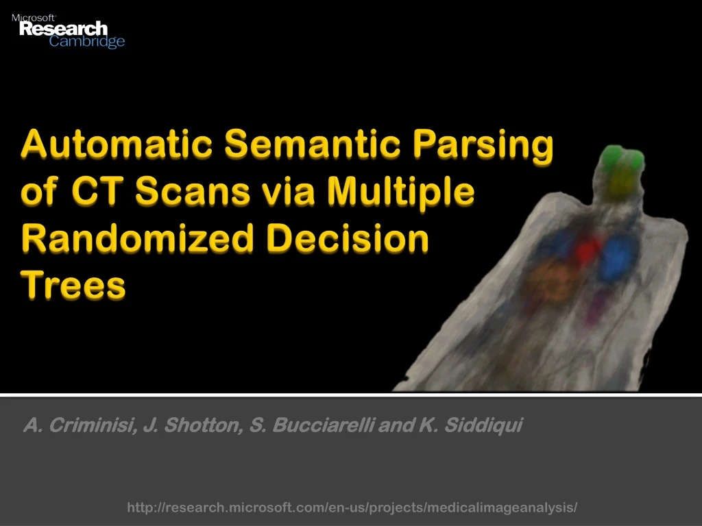
This research project focuses on the detection and localization of anatomical structures in CT scans using multiple randomized decision trees. The aim is to improve visualization, navigation, and initialization for organ-specific processing, as well as enable content-driven image search.

E N D
Automatic Semantic Parsing of CT Scans via Multiple Randomized Decision Trees A. Criminisi, J. Shotton, S. Bucciarelli and K. Siddiqui http://research.microsoft.com/en-us/projects/medicalimageanalysis/
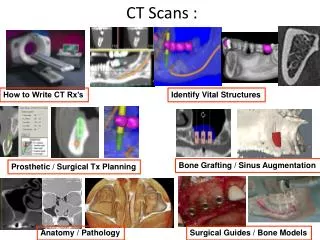
Detection of anatomical structures, why? Detected structures Heart mitral valve left ventricle right ventricle Angio abd. aorta l. renal arteries r. renal arteries Liver possible calcif. Spleen Lungs left right Kidneys left right Spine • One-click visual navigation • Better visualization (class-driven col. transfer functions) • Initialization for organ-specific processing • Content-driven image search Applications One-click visual navigation
Detection of anatomical structures, why? Detected structures Heart mitral valve left ventricle right ventricle Angio abd. aorta l. renal arteries r. renal arteries Liver possible calcif. Spleen Lungs left right Kidneys left right Spine • One-click visual navigation • Better visualization (class-driven col. transfer functions) • Initialization for organ-specific processing • Content-driven image search Applications Setting up the ideal 3D view for diagnosing problems with heart valves is laborious.
Detection of anatomical structures, why? Detected structures Heart mitral valve left ventricle right ventricle Angio abd. aorta l. renal arteries r. renal arteries Liver possible calcif. Spleen Lungs left right Kidneys left right Spine Applications • One-click visual navigation • Better visualization (class-driven col. transfer functions) • Initialization for organ-specific processing • Content-driven image search If we know where the liver is then we can start an automatic process for detecting calcifications.
Detection of anatomical structures, why? Detected structures Heart mitral valve left ventricle right ventricle Angio abd. aorta l. renal arteries r. renal arteries Liver possible calcif. Spleen Lungs left right Kidneys left right Spine Applications • One-click visual navigation • Better visualization (class-driven col. transfer functions) • Initialization for organ-specific processing • Content-driven image search If we know where the liver is then we can start an automatic process for detecting calcifications.
Labelled database:How diverse are the datasets? … Considerable geometric variations. Conventional atlas-based techniques would not work. No contrast agent
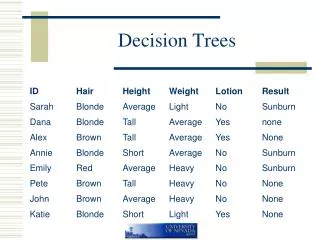
Labelled database: 39 labelled CT volumes Classes = heart, liver, l. kidney, r. kidney, l. lung, r. lung, l. eye, r. eye, head, background Labelling via axis aligned 3D bounding boxes. Positive and negative training examples for organ centres.
Random forest classifier (training) class Training a single tree class class S class S1 S2 class Node optimization function During training each node “sees” only a random subset of all available features Each tree is training independently, using the same procedure
Random forest classifier (testing ) Testing Posterior output of classifier Organ detection Organ localization Using multiple trees has been shown to improve generalization.
Capturing long-range spatial context Context-rich visual features, a 2D illustration Feature response Lots and lots of randomly generated features. Out of those the most discriminative ones are selected automatically during training. Long-range spatial context is captured by the displaced integration regions.
Qualitative evaluation Results of automatic organ detection and localization for three different patients.
Quantitative evaluation (multiple runs on multiple train/test splits) • Our algorithm • Gaussian Mix. Model • Template matching
Future work • More anatomical structures • Hierarchical -> Finer structures • Spatial priors for greater robustness to noise • Larger training database http://research.microsoft.com/en-us/projects/medicalimageanalysis/