Download
1 / 38
380 likes | 552 Vues
Cell and Molec. Extracellular Matrix. Extracellular Matrix. An interconnected network of macromolecules secreted by the cells In most tissues, fibroblast cells are primarily responsible for secreting the extracellular matrix. Examples E.M.

E N D
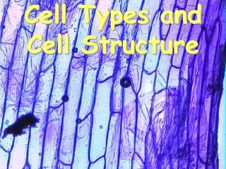
Cell and Molec Extracellular Matrix
Extracellular Matrix • An interconnected network of macromolecules secreted by the cells • In most tissues, fibroblast cells are primarily responsible for secreting the extracellular matrix
Examples E.M. • Bone: Most of bone is a rigid extracellular matrix with only a few cells scattered through it • Cartilage: Almost entirely matrix material • Most glands and blood vessels are surrounded by a gelatinous extracellular matrix which has many cells in it
E.M. provides shape and Support, but also affects: • Cell shape • Cell motility • Growth • Division • Development of specialized cellular characteristics
3 Classes of E.M. Molecules • 1) Glycosaminoglycans & Proteoglycans: gelatinous substance • 2) Structural proteins, (collagens, elastin): give strength and flexibility to the matrix • 3) Adhesive proteins, (fibronectin, laminin): promote attachment of cells to the matrix
Ground Substance of E.M. • Glycosaminoglycans and Proteoglycans • Glycosaminoglycans are polysaccharides • They contain repeating disaccharide containing an amino sugar and at least one negatively charged sulfate or carboxyl group • Since they are hydrophilic (sugar) and (-) charged, they attract H20 and (+) charged molecules, producing a hydrated gelatinous material called the ground substance of the extracellular matrix
Glycosaminoglycans • Chondroitin sulfate • Keretin sulfate • Heparin • Heparin sulfate • Hyaluronate (hyaluronic acid) • Dermatin sulfate
Proteoglycans • Most glycosaminoglycans exist attached to a protein • A proteoglycan is composed of one core protein with multiple attached glycosaminoglycans (may be 95% polysaccharide)
Cartilage, a proteoglycan matrix • Cartilage tissue is composed of dozens of proteoglycan molecules attached to one long hyaluronate backbone • This give cartilage its strength and flexibility properties
Hyaluronic Acid • H.A. is unusual in that it also exists as a free polysaccharide • H.A. is found in high levels in tissues where cells are moving or actively dividing • It is found in the surface of cells which are migrating, but is removed when cells cease to migrate • It is thought to be involved in the movement / migration of cells, possibly by attracting a H2O layer • H.A. also is seen as a “lubricant” in joints between bones
Collagen • Collagen is a group of related proteins (14 types or more). • 30% of the total protein of a vertebrate is collagen • Made of 3 intertwined polypeptide chains: these alpha chains form a triple helix
Collagen • 25% of the amino acids in collagen are glycine • Another 25% are unusual amino acids hydroxy proline and hydroxy lysine • Hydrogen bonding between the OH of the hydroxy proline and the H of glycine gives strength to the triple helix
Collagen • Scurvy, caused by a lack of Vitamin C, is due to a loss of activity of the enzyme which produces hydroxy proline. Therefore, the triple helix is destabilized, resulting in a loss of structure in the connective tissue. Bruising, bleeding, and other connective tissue problems result.
Collagen • More than 14 types of collagen are formed by various combination of chains from at least 20 different genes. • They exist in one of two forms: banded fibrils or unbanded filamentous networks • Larger fibers of collagen form in some tissues, giving strength to tendons, cartilage, etc.
Formation of Collagen • Collagen protein is produced as a precurser chain with extra amino acids at both ends • These extra amino acids are necessary for the triple helix to form • 3 chains form a triple helix procallogen in the ER lumen • The extra A.A.s then prevent the formation of fibrils • The procallogen is secreted from the cell, the extra amino acids are removed (by procallogen peptidases) • The collagen then spontaneously forms fibrils • Crosslinks between lysines and hydroxylysines add strength to the networks/
Other ECM Components • 1)Elastin: Flexibility, glycine and proline rich • 2)Fibronectin: one gene, many forms due to alternative splicing of the RNA • Attach the cell surface to the ECM. • Guide cells during migration (embryonic development, immune response to wounds, etc.) • Different domains interact with different proteins
Other ECM Components • 3) Laminin: in the basal lamina, a thin sheet of ECM separating epithalial cells from the underlying supporting tissues • Surround many nerve, muscle, and fat cells • Provides separation of cell types, influences growth patterns, differentiation, motility • 4) Integrin receptors: transmembrane proteins which bind ECM on the outside of the cell, bind the cytoskeleton on the inside. “Integrate the organization of the cytoskeleton with that of the extracellular matrix”
Glycocalyx • A carbohydrate-rich zone located at the periphery of many animal cells • Involved in cell recognition, adhesion, protection of cell surface, permeability barriers • “attached glycocalyx”: glycoprotein and glycolipid carbohydrates • “unattached glycocalyx”: secreted glycoproteins and proteoglycans (Extracellular matrix)
Cell Recognition and Adhesion • Cells seem able to recognize similar cells and adhere specifically to like cells. • Early experiment with two color sponges: cells dis-associated and allowed to reform: only like cells clumped together
Adhesion Molecules • N-CAM: neural cell adhesion molecule: involved in “linking” of neural cells in development • Appears that N-cam molecules the cell surface interact with N-cams on the next cell • Cadherins: A class of Calcium requiring cell adhesion molecules • Epithelial, Nervous, Placental (E-cadherin, N-cadherin, P-cadherin): specific interaction with the same type of cadherin: nerve cells only bind to nerve cells and so forth.
Carbohydrates and Recognition and Adhesion • Most surface recognition and adherin proteins are glycosylated: it seems that the carbohydrate is important for the recognition and adhesion • Lectins are secreted proteins which can bind (multiple) carbohydrates: presumed to be involved in the adhesion process.
Sialic Acid and Cell Aging • RBCs are removed from circulation by the spleen after about 3-4 months • Glycophorin has the carbohydrate sialic acid at the ends of many carbohydrate chains • Loss of sialic acid from the glycoprotein seems to be part of the way the spleen recognizes “old” cells to be removed
Cell Junctions • Three major types of cellular junctions: • 1) Tight Junctions • 2) Adhesive (Plaque-bearing) junctions • 3) Gap junctions
Tight Junctions • Form permeability barriers across cell layers (such as the lining of the digestive tract) • Also form polarity in cells: prevent diffusion of proteins within the membrane across the junction
Plaque-Bearing Junctions • Provide connections to the cytoskeleton between two adjacent cells • Desmosomes: attach to intermediate filaments: plakoglobin and desmoplakin in plaque • Adherins Junctions: provide attachments to actin filaments: vinculin,( talin in focal adhesions)
Gap Junctions • Gap junctions allow passage of small molecules between adjacent cells • Are dependent on Ca+ concentration (close with higher Ca+) • Made of protein called connexons
Plant Cell Wall • Plant cells are surrounded by a rigid cell wall. • The cell wall, like the extracellular matrix of animal cells, is formed from material secreted by the cell. • Water, gases, ions, and small water soluble molecules such as sugars and amino acids can readily diffuse through the cell wall.
Components of the Cell Wall • Cellulose • Hemicelluloses • Pectins • Extensins • Lignins
Cellulose • The most abundant organic macromolecule on earth. • Unbranched polymer of glucose units linked by beta1,4 linkages. • 50-60 molecules form microfibrils, stabilized by hydrogen bonds between molecules. • Microfibrils are often twisted into ropelike macrofibrils. • Cellulose macrofibrils are as strong as a similar sized piece of steel.
Hemicelluloses • A varied group of polysaccharides. • Each is a long chain of a single type of sugar (glucose or xylose) with short side chains. • The side chains contain several types of sugars: • Hexoses; glucose, galactose, mannose • Pentoses; xylose and arabinose • Hemicelluloses form a coating over the cellulose helping to bond the cellulose fibrils into a rigid network.
Pectins • Pectins are polysaccharides with a backbone of negatively charged galacturonic acid and rhamnose. • Pectin side chains are similar to hemicellulose side chains. • Proteins crosslink the pectin backbone to the hemicelluloses. • Pectin forms a matrix in which the cellulose microfibrils are embedded, and bind adjacent cell walls together. • Pectins trap water, forming a gel like substance which can vary from fluid to rigid, depending on the chemical composition of the pectin: pectin is the gelling agent in jam and jelly.
Extensins • Extensins are glycoproteins: the peptide backbone is rich in serine, hydroxyproline, and lysine. • Lysine is + charged, and causes extensins to bind to the – charged pectins. • Extensins are deposited as a soluble molecule, but become covalently crosslinked to one another and to cellulose, forming a reinforced protein-polysaccharide network.
Lignins • Lignins are insoluble polymers of aromatic alcohols found mainly in woody tissues. • The alcohols are deposited in the cell wall, then covalently crosslinked by the enzyme peroxidase. • This network of lignin accounts for up to 25% of the dry weight of wood, and gives wood much of its strength. • Lignin is second only to cellulose in abundance in the organic realm.
Cell Wall Synthesis • Cell wall components are secreted from the cell. • The layer of the cell wall farthest from the cell is secreted first. • The middle lamella is secreted first. • The primary cell wall is secreted second, while the cells are growing. • The secondary cell wall is secreted by some cells after they have ceased growth.
Middle Lamella • Shared by adjacent cells • Holds the cells togeher.
Primary Cell Wall • 100-200 nm thick • Loosely organized network of cellulose microfibrils, hemicelluloses, pectins, and glycoproteins. • Pectins impart flexability, allowing the cell wall to expand during cell growth. • Cellulose microfibrils are synthesised by enzyme compleses called rosettes, which move across the membrane along the newly forming fibril. • A family of proteins called expansins are important in allowing the cell wall to remain pliable.
Secondary Cell Wall • Some cell types stop cell wall synthesis after forming the primary cell wall. • Many cells form a multilayered secondary cell wall after cell growth has ceased. • The secondary cell wall is composed mainly of cellulose and lignin. • These layers are stiff and strong, giving wood much of its strength. • Cellulose microfibrils in each layer are parallel, and those in adjacent layers are at right angles. • Microtubules in the cell are thought to guide the rosettes in forming this regular arrangement of cellulose microfibrils.
Plasmadesmata • The cell wall poses a barrier to cell cell communication in plants. • One part of the solution to this problem is the use of small water soluble “hormones” which can diffuse through the cell wall material. • A second solution is the formation of cytoplasmic channels through the cell wall of adjacent cells, allowing communication (like gap junctions in animal cell, only much larger). • These openings are called plasmadesmata. • Tubular (membrane?) structures called desmotubules are often associated with the plasmadesmata. • Desmotubules appear to allow continuity of the ER network of adjacent cells.