Download
1 / 24
270 likes | 539 Vues
Charles University Physiology 29/04/09. Activity-Dependent Structural and Functional Plasticity of Astrocyte-Neuron Interactions. By: Luís Carvalho. Astrocytes. Number and structure according with complexity of the organism. Numerous in CNS. One astrocyte contacts 1000s synapses.

E N D
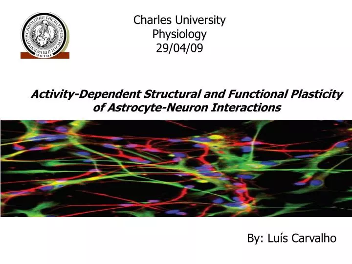
Charles University Physiology 29/04/09 Activity-Dependent Structural and Functional Plasticity of Astrocyte-Neuron Interactions By: Luís Carvalho
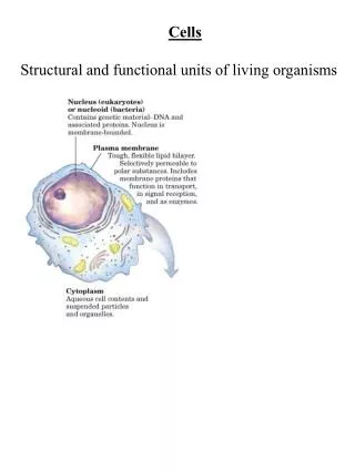
Astrocytes • Number and structure according • with complexity of the organism. • Numerous in CNS. • One astrocyte contacts 1000s • synapses. • Enwraps 4-8 neuronal somata and • 300-600 dendrites. • - Most prominent feature: • Glial fibrillary acidic protein (GFAP). Gray matter - protoplasmic White matter - fibrous
Processing information is not an exclusive property of neurons… • Not only structural and nutritional support. (Brain blood barrier) • Role in regulation of synaptic function. • Can also undergo remodeling. (Plasticity) • - The fine distal processes are interposed between all neuronal elements. • Create a kind of synaptic island defined by its ensheathing processes. V. Parpura, UC-Riverside glial fibrillary acidic protein (GFAP) tagged with antibody.
Tripartite Synapse • Considered a physical barrier to restrict spill over and diffusion of released molecules to ECS. • - Position of relevance to their functions. (Araque et al., TINS 22 (1999))
Astrocyte Role: Metabolic support: provide neurons nutrients such as lactate As Neural activity there is an Energy requirement To solve this… Astrocytic uptake of Glutamate leads to> ADP leads to> Glycolysis within Astrocytic endfeetwhich finally leads to>Lactate delivered to neuron
Regulation of ion concentration in the ECS: • Ex: High number of K+ channels (high permeability). • Transference of K+ to sites of lower accumulation. High levels of K+ in ECS would change neuronal exitability. • Clear neurotransmitters (glutamate and GABA): • Astrocytes have distal processes rich in transporters that remove excess neurotransmitters (especially glutamate) • If Glutamate is not removed: • Diffuses into the ECS. Presynaptic bind and inhibition of its own release. • Influence other synapses - “Intersynaptic cross-talk”
- Secrete large complex substances to the ECS: Important as structural elements and cell to cell communication. • Ex: Promotion of the myelinating activity of oligodendrocytes through release of cytokine leukemia inhibitory factor (LIF). • - Nervous system repair: upon injury to nerve cells within the central nervous system, astrocytes become phagocytic to ingest the injured nerve cells. The astrocytes then fill up the space to form a glial scar, repairing the area and replacing the CNS cells that cannot regenerate.
Vasomodulation: Restrict access of neurosecretory terminals to perivascular basal lamina. (blood flow) • Control the effect of paracrine/autocrine secreted peptides. • Regulate neurosecretion. - Modulation of synaptic transmission
Neuron to Astrocyte Signaling IV. Intracellular levels of Ca2+ rise., free Ca2+ releases other pools of vesicular-bound Ca2+. II. Metabotropic receptors for Glutamate (mGluR) located on astrocyte bind synaptic Glutamate. Subsequent intracellular Phospholipase C release leads to Inositol Triphosphate (InsP3) production. III. Ion channels open, allowing vesicular-bound pools of Ca2+ into the intracellular enviornment. I. Glutamate release from pre-synaptic neuron
Ca2+ increase… • - Can be also caused by increased extracellular K+ levels. • - Modify gene expression and consequent morphological changes. • Cause own release of glutamate. • Further adjacent neuron activation. (not confirmed) Basis of Matainance of microvascular tone
Astrocytes are connected bygap junctions thereby forming a syncytium that is able to propagate signals for large distances Ca2+ Increase cause… • Wave propagation signal • - Mechanism of wave propagation via • release of ATP to ECS > Activates neighboring cells. • - Thigh junctions. Not certain. • Observed only in intense electrical stimulation
Ca2+ Astrocyte Ca2+ Glu Gliotransmission Glutamate: Post synaptic - contribute to network synchronization Pre synaptic - facilitates subsequent glutamate release. Favoring neurotransmission - ionotropic receptors Inhibition - metabotropic receptors
Synchronous Firing Groups - Astrocytic Regulation of Neural Networks
D serine Important intermediary in glutamate neurotransmission together with glutamate ionotropic receptors. Instead of glycine (hippocampus, retina, hypothalamus). TNF - a Promotes the neuronal insertion of AMPA receptors enhancing and maintaining synaptic strength.
ATP and adenosine: ATP - P2Y receptors in astrocytes. Triggers intracellular Ca2+ release and wave propagation. > Glutamate Signal neighboring neurons by pre/post synaptic purinergic receptors. Converted to adenosine by ectonucleotidases in ECS. Suppression of synaptic transmission. A1/A2 receptors activation leads to positive action of K+ channels and negative action of Ca2+ channels.
Astrocytic Mobility - Constantly changing their morphology. - Specially distal processes devoid of GFAP are extremely mobile. (GFAP imunolabelings show even in normal conditions) Examples: Long term potentiation (LTP) Observed in Hippocampus - increase of density and closer apposition to synaptic cleft of potentiate synapses. Astrocyte Remodeling.
Substances like OT are release by neurohypophysis in times of parturition, lactation or chronic dehydration. Astrocytes - significant reduce coverage of OT neurons. Neurons- Somata hypertrophy, branch of dendrites and axons enlarge and ramify. Juxtaposed surfaces and dendrites
Cerebellum Bergmann glia require constant input from their associated synapses to maintain their relationship. AMPA receptors (also for glutamate) + GLuR2 subunit are responsible for Ca2+ permeability. Removal of the subunits reduced permeability and provoked retraction of glia.
Consequences of Extracellular Homeostasis Any structural change in astrocyte environment should affect properties of ECS. Observing on rats. During lactation: Significant reduction of volume fraction and tortuosity (diffusion rate compared with obstacle-free medium) Diffusion became equivalent in all planes (isotropy, instead of anisotropy in normal conditions) Diffusion properties changed as well. Enhance the range of action of molecules.
Neuropathological conditions Epilepsy - acompained by astrocyte hypertrophy and hyperplasia Schizophrenia - astrocytes produce factors like Bornea disease virus Phosphoprotein which have been linked to bahavioral abnormalities in mice. Astrocytes activated by injury - regulation of synaptic activity and strength. Importance in development of inflammatory pain.
End! Thank you for your attention… Bye!