Download
1 / 16
250 likes | 1.24k Vues
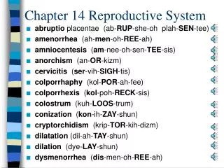
Chapter 16 The Reproductive System. Notice: This presentation contains actual pictures of human reproductive anatomy. Female Reproductive System. Ovaries Duct System - Uterine tubes (fallopian tubes) - Uterus - Vagina External genitalia. Ovarian Follicle Stages.

E N D
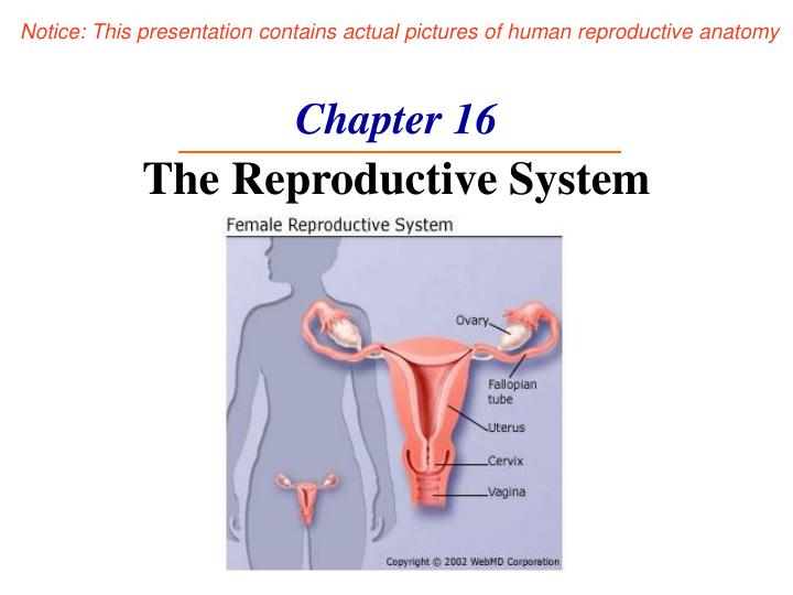
Chapter 16The Reproductive System Notice: This presentation contains actual pictures of human reproductive anatomy
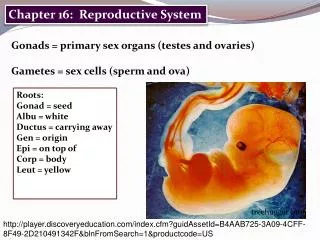
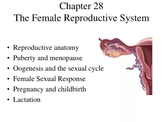
Female Reproductive System • Ovaries • Duct System - Uterine tubes (fallopian tubes) - Uterus - Vagina • External genitalia
Ovarian Follicle Stages • Primary follicle –immature oocyte • Graafian (vesicular) follicle – growing follicle; maturing oocyte • Ovulation – follicle ruptures when egg is mature ~ every 28 days • Ruptured follicle transformed intocorpus luteum
Uterine (Fallopian) Tubes • Receive oocyte • Site of fertilization • Attaches to uterus • Does not attach to ovary • Supported by broad ligament
Uterine Tube Function • Fimbriae – finger-like projections at end; receive oocyte • Cilia inside uterine tube slowly move oocyte towards uterus (takes 3–4 days) • Fertilization occurs inside uterine tube
Uterus • Between bladder and rectum • Hollow organ • Functions • Receives • Retains • Nourishes fertilized egg
Support for the Uterus • Broad ligament – attached to pelvis • Round ligament – anchored interiorly • Uterosacral ligaments – anchored posteriorly
Regions of the Uterus • Body – main portion • Fundus – area where uterine tubes enter • Cervix – narrow outlet that protrudes into the vagina Cat Fundus
Walls of the Uterus • Endometrium - Inner layer - Allows for implantation of a fertilized egg - Sloughs off if no pregnancy occurs (menses) • Myometrium – middle layer of smooth muscle • Serous layer – outer visceral peritoneum
Vagina • From cervix to exterior • Behind bladder; in front of rectum • Birth canal • Receives penis during intercourse • Hymen – partially closes the vagina until it is ruptured
External Genitalia (Vulva) • Mons pubis - Fatty area overlying the pubic symphysis - Covered with pubic hair after puberty • Labia – skin folds - Labia majora - Labia minora
External Genitalia Vestibule • Enclosed by labia majora • Contains opening of urethra and vestibular glands (produce mucus) Clitoris • erectile tissue • Corresponds to penis
Oogenesis • Total supply of eggs are present at birth • Ability to release eggs ~ begins at puberty ~ ends at menopause • Oocytes - matured in developing ovarian follicles
Oogenesis • Oogonia – female stem cells • Mitosis produces primary oocytes • Primary oocytes -surrounded by cells that form primary follicles • Oogonia no longer exist by birth
Oogenesis • Follicle stimulating hormone (FSH) causes some follicles to mature • Meiosis starts inside maturing follicle • Produces secondary oocyte and first polar body • Meiosis is completed after ovulation only if sperm penetrates • Two additional polar bodies are produced