Download
1 / 20
220 likes | 924 Vues
PART 3. The Urinary System. Microscopic Anatomy of the Kidney. Juxtaglomerular apparatus Functions in the regulation of blood pressure Juxtaglomerular cells – secrete renin Macula densa A portion of distal convoluted tubule Tall, closely packed epithelial cells Act as chemoreceptors.

E N D
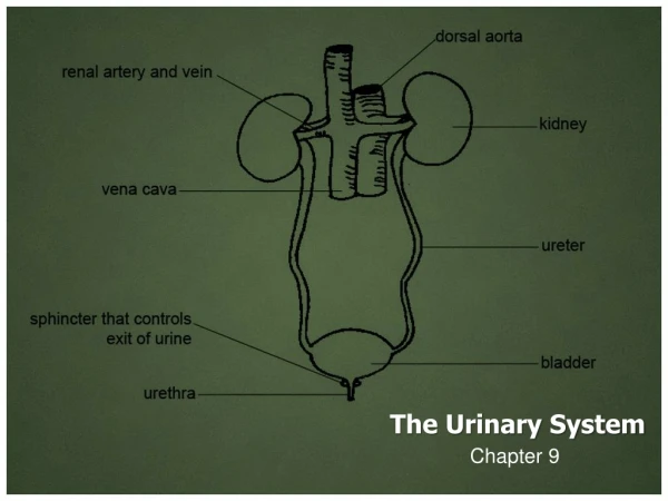
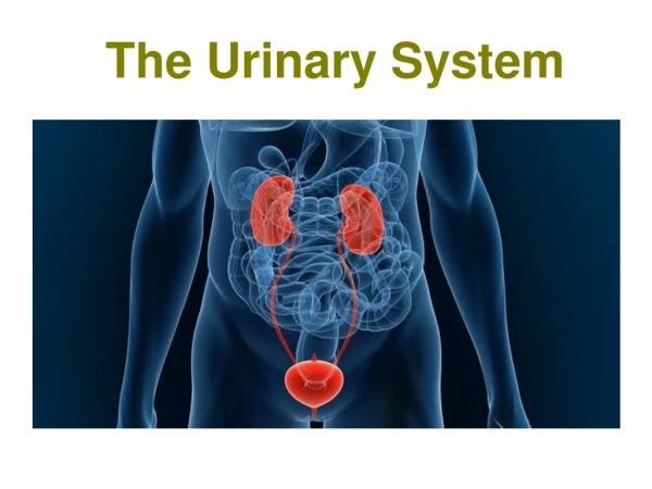
PART 3 The Urinary System
Microscopic Anatomy of the Kidney • Juxtaglomerular apparatus • Functions in the regulation of blood pressure • Juxtaglomerular cells – secrete renin • Macula densa • A portion of distal convoluted tubule • Tall, closely packed epithelial cells • Act as chemoreceptors
Juxtaglomerular Apparatus Figure 23.10
Ureters • Carry urine from the kidneys to the urinary bladder • Oblique entry into bladder prevents backflow of urine • Histology of ureter • Mucosa – transitional epithelium • Muscularis – two layers • Inner longitudinal layer • Outer circular layer • Adventitia – typical connective tissue
Microscopic Structure of the Ureter Figure 23.12
Urinary Bladder • A collapsible muscular sac • Stores and expels urine • Full bladder – spherical • Expands into the abdominal cavity • Empty bladder – lies entirely within the pelvis Figure 23.13
Urinary Bladder • Urachus – closed remnant of the allantois • Prostate gland • In males • Lies directly inferior to the bladder • Surrounds the urethra Figure 23.14
Urinary Bladder • Wall of bladder • Mucosa • Transitional epithelium • Muscular layer • Detrus or muscle • Adventitia
Histology of the Urinary Bladder Figure 23.15a, b
Structure of the Urinary Bladder and Urethra Figure 23.16a
Structure of the Urinary Bladder and Urethra Figure 23.16b
Urethra • Epithelium of urethra • Transitional epithelium • At the proximal end (near the bladder) • Stratified and pseudostratified columnar – mid urethra (in males) • Stratified squamous epithelium • At the distal end (near the urethral opening)
Urethra • Internal urethral sphincter • Involuntary smooth muscle • External urethral sphincter • Voluntarily inhibits urination • Relaxes when one urinates
Urethra • In females • Length of 3–4 cm • In males – 20 cm in length – three named regions • Prostatic urethra • Passes through the prostate gland • Membranous urethra • Through the urogenital diaphragm • Spongy (penile) urethra • Passes through the length of the penis
Micturition Figure 23.17
Disorders of the Urinary System • Urinary tract infections • More common in females • Burning sensation during micturition • Renal calculi • Kidney stones • Bladder cancer • 3% of cancers – more common in men • Kidney cancer • Arises from epithelial cells of uriniferous tubules
The Urinary System Throughout Life • Embryo develops three pairs of kidneys • Pronephros • Mesonephros • Metanephros • Only metanephros persists to become the adult kidneys • Metanephric kidney produces urine by fetal month three • Contributes to the volume of amniotic fluid
Development of the Urinary Organs Figure 23.18a,b
Development of the Urinary Organs Figure 23.18c, d
The Urinary System Throughout Life • Kidney and bladder function declines with advancing age • Nephrons decrease in size and number • Tubules less efficient at secretion and reabsorption • Filtration declines • Recognition of desire to urinate is delayed • Loss of muscle tone in the bladder