Download
1 / 35
390 likes | 1.17k Vues
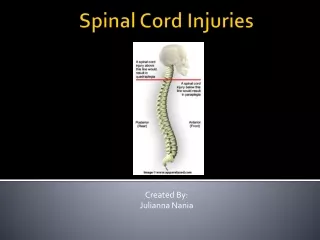
Spinal cord injuries. Overview:. Anatomy of the spinal cord Case presentation Spinal cord injuries Classification Complete and incomplete syndromes Respiratory complications of spinal cord injuries ICU management of spinal cord injuries Pharmacological management. Gross anatomy.

E N D
Overview: • Anatomy of the spinal cord • Case presentation • Spinal cord injuries • Classification • Complete and incomplete syndromes • Respiratory complications of spinal cord injuries • ICU management of spinal cord injuries • Pharmacological management
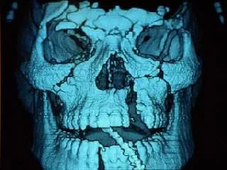
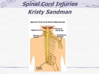
Gross anatomy • Begins at the foramen magnum of the skull, where it is continuous with the medulla oblongata • Cervical enlargement gives rise to the brachial plexus • Lumbar enlargement gives rise to the lumbosacral plexus • Tapers inferiorly to the conus medullaris – from here the filum terminale attaches to the coccyx • Lower end of the spinal cord lies at the lower border of L1 • Vertebral column is much longer than the spinal column, so the cord segments do not correspond numerically to the vertebral bodies
Columns of the spinal cord • Spinal column stabilised by three major ligaments; • Anterior longitudinal ligament • Posterior longitudinal ligament • Ligamentum flavum • Anterior column: Anterior 2/3 vertebral bodies and the anterior ligament • Middle column: Posterior 1/3 vertebral bodies and the posterior ligament • Posterior column: Ligamentum flavum and everything else • Injury involving > one column is considered unstable
Spinal cord structure • Inner core of grey matter, surrounded by an outer covering of white matter • Grey matter is arranged in an ‘H- shape’, with anterior and posterior horns, joined by a thin grey commissure which contains the central canal • The T1-L3 segments also contain a lateral grey horn
Grey matter of the spinal cord • The anterior horn is divided into medial, central and lateral columns • Medial group is present in most segments innervating the skeletal muscles of the neck and trunk • Central group is the smallest and is present in some cervical and lumbosacral segments • Lateral group is present in the cervical and lumbosacral segments innervates the skeletal muscles of the limbs • Posterior horn has four different groups of nerve cells • Substantia gelatinosa group • Nucleus propius • Nucleus dorsalis • Visceral afferent nucleus • Lateral grey horns contain pre-ganglionic sympathetic fibres
Tracts of the spinal cord • Ascending (sensory): • Dorsal (posterior) columns: deep touch, proprioception, vibration • Lateral spinothalamic: pain and temperature • Anterior spinothalamic: light touch • Descending (motor): • Lateral corticospinal: voluntary motor • Anterior corticospinal: voluntary skilled motor • Rubrospinal: control of movement • Vestibulospinal: posture and balance • Tectospinal: reflex postural movements in response to visual stimuli
Spinal cord injuries • Causes: • Majority caused by MVAs; falls; iatrogenic • Mostly young males, but the other demographic includes older people with concurrent degenerative spinal canal narrowing • Frequently associated with other conditions: • Shock syndromes • Other injuries
Say hello to Jim. • 85 year old male who slipped backwards and hit head on towbar behind car • Presented to Tenterfield Hospital, then T/F to Armidale, where CT showed unstable C4/5 # • Transferred to JHH for NSx R/V • Conscious and spontaneously breathing • Hard collar in situ, but poorly fitting • GCS 13: E3V4M6, PEARL • B/G: metastatic prostate ca, HTN, T2DM
Jim’s imaging • Unstable C4/5 fracture
Further examination… • Normal cranial nerve examination • Decreased strength (2/5) and absent reflexes bilaterally in upper limbs • Decreased pain and temperature sensation bilateral hands • Normal strength (5/5), reflexes and sensation in bilateral lower limbs • Developed urinary retention
American Spinal Injury Association Neurological impairment scale
Classifications • Quadriplegia • Injury to the cervical spine, leading to impairment in the arms, trunk, pelvis and legs • Paraplegia • Injury to the thoracic, lumbar or sacral segments, leading to impairment in the trunk, legs and pelvic organs • Complete • No motor or sensory function below the affect level • Incomplete • Some preserved motor or sensory function below the affected level
Complete injury • No voluntary anal contraction • 0/5 distal motor score • 0/2 sensory score • Bulbocavernous reflex present
Incomplete spinal cord injuries • Anterior cord syndrome • Central cord syndrome • Brown-Sequard syndrome • Posterior cord syndrome
Anterior spinal cord syndrome • Injury to the anterior spinal cord caused by either direct compression of the spinal cord, or damage to the anterior spinal artery • Usually from a flexion/compression injury • Bilateral loss of pain, temperature and light touch below the lesion due to disruption of the anterior and lateral spinothalamic tracts • Motor dysfunction due to the disruption of the anterior corticospinal tracts, and damage to the anterior grey horn neurons • Worst prognosis of incomplete SCI • 10-20% chance motor recovery Anterior
Central spinal cord syndrome • Most common incomplete spinal cord injury • Often in the elderly with extension injury mechanisms, due to anterior osteophytes and posterior infolded ligaments • Motor dysfunction due to disruption of the lateral corticospinal tracts and damage to the anterior grey horn neurons • Bladder and bowel involvement • Bilateral loss of pain, temperature and light touch due to disruption of the spinothalamic tracts • Sacral sparing • Good prognosis, but unlikely to regain full function Anterior
Brown-Sequard syndrome • Caused by complete cord hemitransection • Ipsilateral motor dysfunction, with LMN weakness at the level of the injury, and UMN signs below the injury • Ipsilateral proprioception and vibration loss due to posterior column damage • Contralateral pain and temperature loss 2-3 segments below the lesion due to disruption of the spinothalamic tracts • Good prognosis
Posterior cord syndrome • Rare syndrome • Most commonly caused by vascular compromise, with occlusion to the posterior spinal artery • Sensory dysfunction with ipsilateral loss of proprioception and vibration, and preservation of pain and temperature Anterior
Cauda equina syndrome • Caused by damage to the cauda equina, a collection of S1-L5 nerves • Technically a peripheral nerve lesion, so will cause lower motor neuron signs • Presentation: • Saddle anaesthesia, bilateral lower limb sensorimotor loss and pain, bowel and bladder symptoms (especially urinary retention) • Absent or reduced lower limb reflexes, decreased rectal tone • MRI best to evaluate nerve compression • Needs urgent surgical decompression within 48 hours
Jim’s progress notes… • Admitted under NSx • Few episodes of vomiting on ward, during which he likely aspirated • RRT on ward for respiratory arrest – intubated and T/F to ICU • Some more stuff happened…. • Improved and ready to trial extubation…. • Unfortunately, he failed extubation due to hypoxia • Why?
SCI effects on breathing • Loss of intercostal function: • Failure of AP expansion of the ribcage • Chest wall sucked in during diaphragmatic contraction • Loss of lower thoracic segment innervation: • Diaphragm starts in a flatter position, which decreases contraction pressure • Loss of abdominal muscle tone: • As the diaphragm flattens, abdominal contents are pushed outwards and the lower ribcage is pulled inwards, causing paradoxical see-saw breathing • Diaphragm is pulled down by the weight of the abdomen • Inefficient, rapid, shallow breathing results, with more dead space ventilation • Abdominal muscle weakness results in decreased ability to cough and clear secretions
Respiratory management • Airway management • Physiotherapy • Posture • Mucolytics • Abdominal binding • Monitor for infection • Bronchoscopy
Longer term respiratory care • Tracheostomy • More comfortable; minimise laryngeal damage; less dead space compared to ETT; associated with fewer respiratory infections • Weaning from ventilation • Portex sprints are as effective or better than PS weaning, with both superior to SIMV weaning
Cardiovascular complications after SCI • Neurogenic shock • Occurs with lesions above T6 due to loss of sympathetic tone and unopposed parasympathetic tone • Vasodilation and hypotension; bradycardia • Thromboembolism • Due to immobility and venous stasis • Sympathetic hyperreflexia • Unopposed sympathetic tone below the level of injury, triggered by sensory stimuli
Gastrointestinal complications after SCI • Delayed gastric emptying and ileus • Common and may last 2-3 weeks • Aperients, early feeding, NGT, prokinetic agents • Gastric stress ulceration • PPI prophylaxis • Constipation
Metabolic system considerations • Temperature regulation • Hypothermic due to vasodilation • Hyperthermic due to inability to sweat below level of injury • Hyperglycaemia • Common due to stress response • Worsens ischaemic neurological injury
Pharmacological treatment of SCI • Steroids • Previously, high dose methylprednisone was standard of care for SCI • Since shown to significantly increase mortality in patients, compared to placebo • NOGO-A antibody • NOGO-A is an inhibitory molecule that prevents neuronal plasticity and axonal regeneration • Current clinical trial to determine effects of an intrathecal infusion of NOGO-A antibody
References • Snell. Clinical neuroanatomy. 7th Ed. (2010). Lippincott Williams and Williams: Philadelphia • J Patten. Neurological differential diagnosis. 2nd Ed. (1996). Springer: London • M Denton, J McKinlay. Cervical cord injury and critical care. Continuing education in Anaesthesia and Critical Care. (2009). Vol 9: No. 3 • Stahel et al. Management strategies for acute spinal cord injury: current options and future perspectives. Current Opinion Critical Care. (2012). 18:651-660 • A Neill. Basic neuroanatomy for the critically ill. SMACC. http://smacc.net.au/2013/02/basic-neuroanatomy-for-the-critically-ill/ • C Wheeless. Anterior cord syndrome. Wheeless’ textbook of orthopaedics..Lastupdated: 25/4/12. http://www.wheelessonline.com/ortho/anterior_cord_syndrome Date accessed: 30/6/13 • S Hishmeh. Posterior cord syndrome. Orthopaedics:one. Last updated: 22/6/09. http://www.orthopaedicsone.com/display/Main/Posterior+cord+syndrome Date accessed: 30/6/13 • D Moore. Spinal cord injuries. Ortho bullets. Last updated: 20/5/13. http://www.orthobullets.com/spine/2006/spinal-cord-injuries Date accessed: 30/6/13