Download
1 / 43
430 likes | 444 Vues
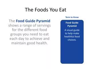
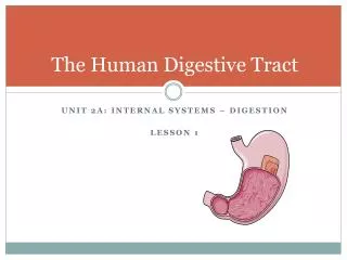
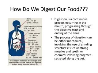
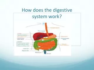
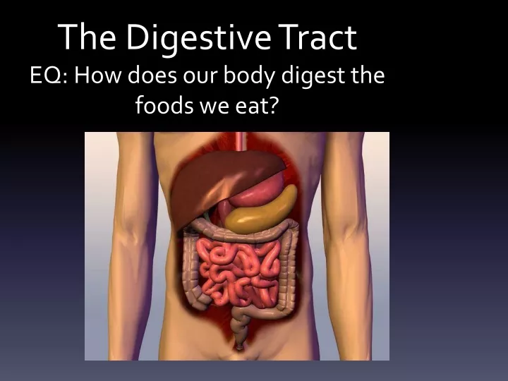
The Digestive Tract EQ: How does our body digest the foods we eat?. Root Words . The GI tract (gastrointestinal tract) The muscular alimentary canal Mouth Pharynx Esophagus Stomach Small intestine Large intestine Anus The accessory digestive organs

E N D
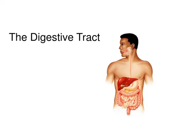
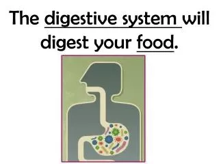
The Digestive TractEQ: How does our body digest the foods we eat?
The GI tract(gastrointestinal tract) The muscular alimentary canal • Mouth • Pharynx • Esophagus • Stomach • Small intestine • Large intestine • Anus • The accessory digestive organs Supply secretions contributing to the breakdown of food • Teeth & tongue • Salivary glands • Gallbladder • Liver • Pancreas
The Digestive Process • Ingestion • Taking in food through the mouth • Propulsion (movement of food) • Swallowing • Peristalsis – propulsion by alternate contraction &relaxation • Mechanical digestion • Chewing • Churning in stomach • Mixing by segmentation • Chemical digestion • By secreted enzymes: see later • Absorption • Transport of digested end products into blood and lymph in wall of canal • Defecation • Elimination of indigestible substances from body as feces
Chemical digestion • Complex food molecules broken down into chemical building blocks • Carried out by enzymes secreted by digestive glands into alimentary canal
Histology of alimentary canal wallSame four layers from esophagus to anal canal • Mucosa • Submucosa • Muscularis externa • Serosa from lumen (inside) out
Inner layer: the mucosa*(mucous membrane) Has folds called Lumens, which help increase absorption area. *
Second layer: the submucosa* • Connective tissue containing major blood vessels and Nerves • Nourishes surrounding tissue *
Muscular Layer Two layers of smooth muscle responsible for movement of material • Inner circular layer • Squeezes • Outer longitudinal layer: • shortens gut *
Serosa • Protect underlying tissue and secretes fluid which lubricates outer surface • Allows abdominal cavity to slide freely *
Omentum: Flap of tissue that hangs from stomach for insulation, protection, and wound isolation
The Mouth • Mouth = oral cavity • Lining: thick stratified squamous epithelium • Lips- orbicularis oris muscle • Cheeks – buccinator muscle • Teeth
Tongue • Mostly muscles • Grip and reposition food • Forms “bolus” of food (lump) • Help in swallowing • Speech – help form some consonants
Teeth • Called “dentition” • Teeth live in sockets (alveoli) in the gum-covered margins of the mandible and maxilla • Chewing: raising and lowering the mandible and moving it from side to side while tongue positions food between teeth
Teeth • Two sets • Primary or deciduous • “Baby” teeth • Start at 6 months • 20 are out by about 2 years • Fall out between 2-6 years • Permanent: 32 total • All but 3rd set of molars by end of adolescence • 3rd set = “wisdom teeth” • Variable • Some can be “impacted” (imbedded in bone)
Teeth are classified according to shape and function • Incisors: chisel-shaped for chopping off pieces • Canines: cone shaped to tear and pierce • Premolars (bicuspids) and • Molars - broad crowns with 4-5 rounded cusps for grinding incisor canine premolar molar Cusps are surface bumps
Salivary glands(tuboalveolar glands) • Parotid • Largest of salivary glands • Between skin of cheek • Submandibular • Located in the floor of lower jaw • Sublingual • Smallest salivary gland • Floor of jaw, inferior to tongue Saliva: mixture of water, ions, mucus, enzymes keep mouth moist dissolves food so can be tasted moistens food starts enzymatic digestion buffers acid antibacterial and antiviral
Pharynx ___oropharynx • Muscular walls allows for swallowing ___laryngopharynx * * *
Esophagus • Passage from pharynx to stomach • Contains muscles to move food downward Esophagus___________ *
Microscopic anatomy of esophagus Contains all 4 layers (see right) • Epithelium: nonkeratinized stratified squamous epithelium • At GE junction – thin simple columnar epithelium • Mucus glands in wall • Muscle (muscularis externa) changes as it goes down • Superior 1/3 of esophagus: skeletal muscle (like pharynx) • Middle 1/3 mixture of skeletal and smooth muscle • Inferior 1/3 smooth muscle (as in stomach and intestines) • When empty, mucosa and submucosa lie in longitudinal folds
Stomach • Temporary storage and mixing – 4 hours • Into “chyme” • Starts food breakdown • Pepsin • HCl (hydrochloric acid) helps kill bacteria • Most nutrients wait until get to small intestine to be absorbed; exceptions are: • Water, electrolytes, some drugs like aspirin and alcohol (absorbed through stomach)
Small intestine • Longest part of alimentary canal (2.7-5 m) • Most enzymatic digestion occurs here • Most enzymes secreted by pancreas, not small intestine • 3-6 hour process Small intestine___________
Small intestine designed for absorption • Villi (fingerlike projections) 1 mm high – simple columnar epithelium: velvety Absorptivie cell with microvilli to increase surface area & many mitochondria: nutrient uptake is energy-demanding * Lacteal*: network of blood and lymph capillaries -Carbs and proteins into blood to liver via hepatic portal vein -Fat into lymph: fat-soluble toxins e.g. pesticides circulate systemically before going to liver for detoxification
Intestinal flora– the permanent normal bacteria • Manufacture some vitamins, e.g. K, which get absorbed -have many mitochondria: nutrient uptake is energy-demanding Duodenal glands* * • Mucus to counteract acidity from stomach • Hormones: • Cholecystokinin (stimulates GB to release stored bile, also pancreas) • Secretin (stimulates pancreatic ducts to release acid neutralizer) * -produce mucus
Large intestine Digested residue reaches it Main function: to absorb water and electrolytes
Rectum • In pelvis • No teniae • Strong longitudinal muscle layer • Has valves * * *
Defecation • Triggered by stretching of wall, mediated by spinal cord parasympathetic reflex
Histology – large intestine • No villi • Fewer nutrients absorbed • A lot of goblet cells for mucus • Lubricates stool
The Liver • Largest gland in the body (about 3 pounds) • Over 500 functions • Main job is to filter blood coming from digestive tract
posterior anterior
Just some of the liver’s repertoire • Produces bile • Picks up glucose from blood • Stores glucose as glycogen • Processes fats and amino acids • Stores some vitamins • Detoxifies poisons and drugs • Makes the blood proteins
Gallbladder* • Bile is produced in the liver • Bile is stored in the gallbladder • Bile is excreted into the small intestine when needed to dissolve fat and cholesterol *
Pancreatic endocrine function(hormones released into blood) • Islets of Langerhans (AKA “islet cells”) are the hormone secreting cells • Insulin (from beta cells) • Lowers blood glucose (sugar) • Glucagon (from from alpha cells) • Raises blood glucose (sugar) (more later)
LEFT SIDE ACTIVITY TRACE THE STEPS THAT FOOD TAKES THROUGH YOUR BODY, FROM MOUTH TO ANUS.