Download
1 / 66
660 likes | 846 Vues
Haemostatic Challenges of Haemopoietic Stem Cell Transplantation. Omolade Awodu FMCPath July 2013. O bjectives. Factors contributing to haemostatic challenges in HSCT Preventing haemostatic challenges Tackling Haemostatic Challenges. 2. My Talk. Overview of normal haemostasis

E N D
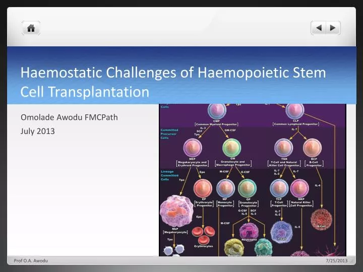
Haemostatic Challenges of Haemopoietic Stem Cell Transplantation Omolade Awodu FMCPath July 2013 Prof O.A. Awodu
Objectives • Factors contributing to haemostatic challenges in HSCT • Preventing haemostatic challenges • Tackling Haemostatic Challenges Prof O.A. Awodu 2
My Talk • Overview of normal haemostasis • Types of haemostatic challenges • Preventing haemostaticinbalance? • Management of haemostatic challenges resulting from HSCT Prof O.A. Awodu
HEMOSTASIS Definition • Hemostasis: drives from the Greek meaning “The stoppage of blood flow”. • There are threehaemostatic components: • The extra-vascular(The tissues surrounding blood vessels) involved in Hemostasis when local vessel is injured. • 2- Thevascular(The blood vessels) it depends on the size, amount, of smooth muscle within their walls and integrity of the endothelial cell lining. • 3- Theintra-vascular (The platelets and plasma proteins that circulate within the blood vessels). Prof O.A. Awodu
NORMAL HAEMOSTASIS: It is a complex process depending on interaction between vessel wall, platelets and coagulation factors. Prof O.A. Awodu
In physiological conditions, the process of thrombin formation and dissolution is maintained in a delicate balance Prof O.A. Awodu
BV Injury Haemostasis: Tissue Factor Neural Coagulation Activation Blood Vessel Constriction Platelet Activation Primary hemostatic plug Reduced Blood flow Plt-Fusion Thromibn, Fibrin Stable Hemostatic Plug Prof O.A. Awodu
HEMOSTATIC SYSTEM INTRINSIC PATHWAY EXTRINSIC PATHWAY COMMON PATHWAY Thrombin Fibrinogen Fibrin PLATELETS CLOT FIBRINOLYSIS TISSUE FACTOR COLLAGEN Prof O.A. Awodu VESSEL WALL
NORMAL HAEMOSTASIS: FIBRINOLYSIS • It helps to restore vessel potency by dissolution of fibrin. • Normal plasma protein Plasminogen (formed in the liver) is converted by Plasminogen activators derived from plasma endothelial cells, platelets, leukocytes and urine into plasmin. Prof O.A. Awodu
FVa Protein S FVIIIa Activation of protein C Activated protein C Thrombomodulin Thrombomodulin PAI-1 TAFI Protein C Thrombin EPCR Endothelial cell Prof O.A. Awodu
My Talk • Overview of normal haemostasis • Types of haemostatic challenges • Preventing haemostaticinbalance? • Management of haemostatic challenges resulting from HSCT Prof O.A. Awodu
Haemostatic Challenges in HSCT Prof O.A. Awodu
Definition • Haematopoietic stem cell transplantation (HSCT) is the transfer of multipotenthaematopoietic stem cells from a healthy donor after myeloablative and non myeloablative conditioning • It is currently the only curative option for many malig- nant and non-malignant haematological diseases. Peripheral blood, bone marrow or umbilical cord are used as stem cell source. Prof O.A. Awodu
Haemostatic challenges Prof O.A. Awodu
Prothrombotic factors • Endothelial injury Prof O.A. Awodu
Causes of endothelial injury in HSCT Prof O.A. Awodu
endothelium Prof O.A. Awodu
In Case if there is an Endothelial Injury(Bleeding must be prevented at site of injury) Prof O.A. Awodu
Other prothrombotic factors Prof O.A. Awodu
Sparse Data on alterations of procoagulant, anticoagulant and fibrinolytic factors in the early phase after HSCT Prof O.A. Awodu
Decrease levels of natural anticoagulants: • protein C • antithrombin • factors V and X and plasminogen • Gordon B, Haire W, Kessinger A et al. High frequen-cy of antithrombin 3 and protein C deficiency following autologous bone marrow transplantation for lymphoma. Bone Marrow Transplant 1991; 8: 497–502. • Collins P, Roderick A, O'Brien D et al. Factor VIIa and other haemostatic variables following bone marrow transplantation. ThrombHaemost 1994; 72: 28–32. Prof O.A. Awodu
VTE, catheter-related thrombosis • Studies have shown that at about six months after transplantation, the incidence of symptomatic venous thromboembolism (VTE) is about 4.9%. • The majority of events were catheter- related VTEs (3.6%), • lower extremity DVT (0.7%) • pulmonary embolism (0.6%) • Gerber DE, Segal JB, Levy MY et al. The incidence of and risk factors for venous thromboembolism (VTE) and bleeding among 1514 patients under- going hematopoietic stem cell transplantation: im- plications for VTE prevention. Blood 2008; 112: 504–510. Prof O.A. Awodu
Risk factors for VTE • The main risk factors predisposing to VTE were: • History of prior VTE (odds ratio 2.9) • Development of graft-versus-host disease (OR 2.4). • Allogeneic transplantation have higher rates of VTE compared to those treated with autologous transplantation Prof O.A. Awodu
VTE • within the allogeneic population, development of GVHD is consistently associated with an increased risk of VTE. • Increase thrombin generation and decrease PAI-1 have been shown to correlate with onset of GVHD • Pinomaki A, Volin L, Joutsi-Korhonen L et al. Bone Marrow Transplant 2010; 45: 730–737. Prof O.A. Awodu
Polymorphisms in coagulation proteins such as the factor V G1691A mutation (fac- tor V Leiden) or the prothrombin G20210A mutation are well-established risk factors for VTE in the general population. • Limited studies on their effects in HSCT patients Prof O.A. Awodu
Treatment and prevention of VTE • Exact role of thromboprophylaxis in catheter related VTE not clear • A single arm trial with minidose warfarin (1 mg/d fixed dose if platelet count >50 x 10 9/l has been reported to produce a comparatively low rate of VTE Prof O.A. Awodu
VTE treatment in HSCT • Treatment of established VTE after HSCT is as for thromboembolic events outside the HSCT setting. • Special precaution must be taken because of the increased risk of bleeding resulting from thrombocytopenia and mucositisin the immediate post-transplant period, this may necesscitatedose reduction of the anti- coagulants Prof O.A. Awodu
Patients must be monitored intensively. • Haemostaticsupport with platelet concentrates must be readily available Prof O.A. Awodu
Pathogenesis of VOD Prof O.A. Awodu
Diagnostic Criteria for VOD Seattle Baltimore Hyperbilirubinaemia and two additional factors must be present within 100 days of transplantation) ● hyperbilirubinemia (total serum bilirubin >2 mg/dl)● Hepatomegaly ● Right upper quadrant pain of liver origin● Sudden weight gain (>5% of baseline body weight) Jones RJ, Lee KS, Beschorner WE et al Transplantation 1987 • At least two factors must be present within 20 days after transplantation) ● Hyperbilirubinaemia(total serum bilirubin >2 mg/dl)● Hepatomegaly or upper right quadrant pain of liver origin • ● Sudden weight gain (>2% of baseline body weight) • McDonald GB, Sharma P, Matthews DE et al .Hepatology 1984 Prof O.A. Awodu
Diagnosis confirmed by histology • Microthrombosesand fibrin deposition in hepatic central venules • Hepatic congestion, and signs of portal hypertension in the absence of in- flammatory infiltrates. • Reversal of the blood flow in hepatic veins documented by duplex ultrasound analysis also supports the diagnosis. Prof O.A. Awodu
Plasmatic markers of coagulation are all up regulated in VOD • Thrombomodulin, P- and E-selectins, • Tissue factor pathway inhibitor (TFPI), • Soluble tissue factor, • Plasminogen activator inhibitor (PAI-1) have all been shown to be up-regulated during VOD. • An elevated PAI-1 level has also been advocated as a diagnostic and prognostic marker for VOD Prof O.A. Awodu
The frequency of VOD is in the range of 10% after allogeneic stem cell transplant Prof O.A. Awodu
Risk factors for VOD • Pre-existing hepatic damage ● previous high-dose chemotherapy ● Previous abdominal irradiation ● Donor-recipient HLA disparity and ● Female gender. Salat C, Holler E, Kolb HJ et al. Blood 1997 Prof O.A. Awodu
Treatment of VOD • Mild VOD may resolve spontaneously • the prognosis of patients with advanced stages is grim. • Presently no treatment with proven efficacy for VOD • Treatment is mainly supportive Prof O.A. Awodu
Defibrotide, a polydisperse mixture of single- stranded oligonucleotides with antithrombotic and fibrinolytic effects on the micro- vascular endothelium is increasingly being used to treat VOD. Prof O.A. Awodu
It binds to microvascular endothelium via adenosine receptors, modulates platelet activity by enhancing levels of endogenous prostaglandins and thrombomodulin, and promotes fibrinolysis via up- regulation of TFPI and tissue plasminogen activator (t-PA). Prof O.A. Awodu
In clinical phase II studies, defibrotide showed remission rates in patients with VOD in the range of 40%. Importantly, responses occurred in the absence of severe hemorrhage or other toxicity. Large phase III trials are currently being conducted. • Richardson PG, Murakami C, Jin Z et al. Blood 2002 Prof O.A. Awodu
Transplant-associated thrombotic microangiopathy(TA-TAM , TAM) • It occurs in 5–10% of patients treated with allogeneic transplantation • Mortality rate in excess of 50%. Prof O.A. Awodu
Thrombocytopenia, schistocytes, increase LDH, bilirubin and reticulocytes as signs of hemolysis, kidney failure, and neurologic abnormalities which are features of TTP are also seen in TA-TAM. Prof O.A. Awodu
TAM mainly results from endothelial injury and not deficiency of ADAMTS-13 • TAM usually develops within the first 100 days after transplantation. Prof O.A. Awodu
Risk factors for the development of TAM ● Fungal or viral infection● Presence of GvHD● Female sex● Unrelated or HLA-mismatched donor grafts,● the use of calcineurininhibitors such as cyclosporine Prof O.A. Awodu
In Case if there is an Endothelial Injury(Bleeding must be prevented at site of injury) Prof O.A. Awodu
Increased percentage of schistocytes in peripheral blood smear (> 4%) ● De novo, prolonged or progressive thrombocytopenia • (< 50 G/l or >50% decrease from previous counts) ● Sudden and persistent increase in lactate dehydrogenase ● Decrease in haemoglobin concentration or increased red blood cell requirements ● Decrease in serum haptoglobin concentration Concensus Criteria for diagnosis of TA-TAM International Working Group Ruutu T, Barosi G, Benjamin RJ et al. Haematologica 2007 Prof O.A. Awodu
RBC fragmentation and ≥ 2 schistocytes per high-power field on peripheral smear • ● concurrent increased serum lactate dehydrogenase above institutional baseline • ● concurrent renal* and/or neurologic dysfunction without other explanations • ● negative direct and indirect Coombs test results Diagnostic criteria BMT CTN Toxicity Committee Ho VT, Cutler C, Carter S et al. Biol Blood Marrow Transplant 2005; Prof O.A. Awodu
Treatment of TA-TAM • No standard treatment for TAM is known. • Plasma exchange is not generally recommended in the treatment of TAM, anecdotal evidence suggests that it may benefit a subgroup of patients. • The demonstration of antibodies against complement factor H in patients with TAM responsive to plasma exchange might pave the way for a more differentiated use of this treatment option in selected patients in the future. • Laskin BL, Goebel J, Davies SM, Jodele S. Blood 2011 Prof O.A. Awodu
Treatment of TA-TAM • Response to the B-cell depleting anti-CD20 antibody rituximab have been reported in some small case series. • Modification of the immunosuppressive regimen is now frequently proposed as the first treatment step: as inhibitors of both calcineurinhave been implicated in the development of TAM • Trials with eculizumab – a monoclonal antibody against the complement protein C5 producing excellent results in atypical hemolytic uremic syndrome – are ongoing. • Au WY, Ma ES, Lee TL et al.Br J Hae- matol2007 • Nurnberger J, Philipp T, Witzke O et al. N Engl J Med 2009 Prof O.A. Awodu
Bleeding complications • Acute bleeding is associated with increased morbidity and mortality, and is a frequent complication after both allogeneic and autologous HSCT • The risk of clinically relevant bleeding is at least 10-fold higher in a transplant population compared to general medical oncology patients under- going chemotherapy • Holler E, Kolb HJ, Greinix H et al. Bone Marrow Transplant 2009 Prof O.A. Awodu
Incidence • In the so far largest cohort study published by Gerber and co-workers 230 of 1514 patients (15.4%) undergoing HSCT experienced clinically significant bleeding complications. • The majority of the bleeding events (39%) occurred in the gastrointestinal tract, followed by genitourinary (23%) and pulmonary bleedings (17%), and haemorrhage of the central nervous system (10%). • Twenty –four % of these events were fatal, 3.6% of the HSCT patients in the cohort died from bleeding. Other authors reported comparable or slightly lower mortality rates from bleeding complications Prof O.A. Awodu