Download

1 / 36
390 likes | 788 Vues
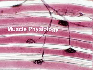
Muscle Physiology. Part II. Types of Muscle. * Voluntary Muscle (Skeletal) Skeletal muscles are usually attached to bone by tendon and moves the skeleton. Skeletal muscle also called striated muscle because of its appearance. * Involuntary Muscle

E N D
Muscle Physiology Part II
Types of Muscle * Voluntary Muscle (Skeletal) Skeletal muscles are usually attached to bone by tendon and moves the skeleton. Skeletal muscle also called striated muscle because of its appearance. * Involuntary Muscle Smooth muscle of the viscera (e.g., in walls of blood vessels, intestine, and other ‘hollow’ structures and organs in the blood. * Cardiac muscle: muscles forming the heart chambers.
Function of the skeletal muscle * Motion: attached to bones and hence responsible voluntary body movement. * Maintenance of posture: Co-ordination of joint movement to maintain the body in either standing, sitting……etc. * Heat production and glycogen storage site.
Function of Smooth Muscle * Regulates the flow of blood in the arteries. * Moves the food along through the gastrointestinal tract. * Expels urine from the urinary bladder. * Regulates the flow of air through the lungs. * Send the babies out into the world from the uterus (uterine contraction).
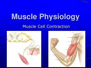
Muscle Responses * Skeletal muscles will not contract unless stimulated by neurons. * Smooth and cardiac muscles will contract without nervous stimulation but their contraction can be influenced by the nervous system. * The nervous and muscle systems are closely interconnected.
Characteristic of muscle * Excitability – responds to stimuli (e.g. nervous impulses). * Contractility – able to shorten in length. * Extensibility – stretches when pulled. * Elasticity – tends to return to original shape & length after contraction or extension.
Neuromuscular (myoneural) Junctions Transmission of impulses from nerve ending to skeletal muscle fibers. The skeletal muscle fibers are innervated by large myelinated nerve fibers that originate from large motoneurons in the anterior horns of the spinal cord. Each nerve fiber, after entering the muscle belly, normally braches and stimulates from three to several hundred skeletal muscle fibers. Each nerve ending makes a junction, called the neuromuscular junction, with the muscle fiber near its midpoint. The action potential initiated in the muscle fiber by the nerve signal travels in both direction toward the muscle fiber ends.
Thick myofilaments * Thick myofilaments are composed of a protein called myosin. Each myosin molecule has a tail which forms the core of the thick myofilament plus a head that projects out from the core of the filament. These myosin heads are also commonly referred to as cross-bridges.
Myosin Head It has several important characteristics: * It has ATP-binding sites into which fit molecules of ATP. ATP represents potential energy. * It has actin-binging sites into which fit molecules of actin. Actin is part of the thin filament. * It has a “hinge” at the point where it leaves the core of the thick myofilament. This allows the head to swivel back and forth, and the “swiveling” is actually the cause of muscle contraction.
Actin filament The actin filament is composed of three protein components: actin, tropomyosin, and troponin. 1. The backbone of the actin filament is a double-stranded F-actin protein. Each strand of the double F-actin helix is composed of G-actin molecules. Attached to each one of the G-actin molecule is one molecule of ADP. 2. Tropomyosin molecules: these molecules are wrapped spirally around the sides of the F-actin helix. In the resting, the tropomyosin molecules lie on top of the active sites of the actin strands.
3. Troponin molecules: these are complexes of three loosely bound protein subunits, each of which plays a specific role in controlling muscle contraction. * Troponin I has strong affinity for actin. * Troponin T has strong affinity for tropomyosin. * Troponin C has strong affinity for calcium ions.
General mechanism of muscle contraction 1. An action potential travels along a motor nerve to its endings on muscle fibers. 2. At each ending, the nerve secretes a small amount of the neurotransmitter substance called acetylcholine. 3. The acetylcholine acts on a local area of the muscle fiber membrane to open multiple “acetylcholine-gated” channels through protein molecules floating in the membrane. 4. Opening of the acetylcholine-gated channels allows large quantities of sodium ions to diffuse to the anterior of the muscle membrane. This initiates an action potential at the membrane. 5. The action potential travels along the muscle fiber membrane in the same way that action potentials travel along nerve fiber membranes.
6. The action potential depolarizes the muscle membrane, and much of the action potential electricity flows through the center of the muscle fiber. Here it causes the sarcoplasmic reticulum to release large quantities of calcium ions that have been stored within this reticulum. 7. The calcium ions initiate attractive forces between the actin and myosin filament, causing them to slide alongside each other, which is the contractile process. 8. After a fraction of a second, the calcium ions are pumped back into the sarcoplasmic reticulum by a Ca⁺⁺ membrane pump, and they remain stored in the reticulum until a new muscle action potential comes along; this removal of calcium ions from the myofibrils causes the contraction to cease.
ATP as the source of energy for contraction 1. Before contraction begins, the head of the cross-bridges bind with ATP. The ATPase activity of the myosin head immediately cleaves the ATP but leaves the cleavage products, ADP plus phosphate ion, bound to the head. In this state, the conformation of the head is such that it extends toward the actin filament but is not yet attached to the actin. 2. When the troponin-tropomyosin complex binds with calcium ions, active sites on the actin filament are uncovered, and the myosin heads then bind with these.
3. The bond between the head of the cross-bridge and the active site of the actin filament causes a conformational change in the head, prompting the head to tilt toward the arm of the cross-bridge. 4. Once the head of the cross-bridge tilts, this allows release of the ADP and phosphate ion that were previously attached to the head. At the site of release of the ADP, a new molecule of ATP binds. This binding of new ATP causes detachment of the head from the actin. 5. After the head has detached from the actin, the new molecule of ATP is cleaved to begin the next cycle, leading to a new power stroke.
Muscle relaxation • * Skeletal muscle relaxes when the nervous impulse stop. * In absence of impulse the membrane of the sarcoplasmic reticulum is no longer permeable to calcium( i.e., no impulse means that the calcium gate close). So, calcium no longer diffuse out. The calcium pump in the membrane will now transport the calcium back into the SR. As this occurs, calcium ions leave the binding sites on the troponin molecule. Without calcium, troponin returns to its original shape and position as does the attached tropomyosin. This means that tropomyosin is now back in position, in contact with the myosin head. So, the myosin head is no longer in contact with Actin, therefore, the muscle stops contracting (i.e., relaxes).
* Calcium is the “switch“ that turns muscle “on and off“ (contracting and relaxing). * when a muscle is used for an extended period, ATP supplies can diminish. As ATP concentration in a muscle declines, the myosin head remain bound to actin and can no longer swivel. This decline calcium in ATP levels in a muscle causes muscle fatigue. Even though calcium is still present. (and a nervous impulse is being transmitted to the muscle), contraction ( or at least a strong contraction) is not possible.
Types of muscle contraction 1. Isotonic – tension for force generated by the muscle is greater than the load and the muscle shortens. 2. Isometric – load is greater than tension or force generated by the muscle and the muscle does not shorten.
Muscle graded response * An important characteristic of skeletal muscle is its ability to contact to vary degree. A muscle, like the biceps, contracts with varying degrees of force depending on the circumstance (this is also referred to as graded response). Muscle do this by a process called summation, specifically by motor unit summation and wave summation.
Motor unit summation * The degree of contraction of a skeletal muscle is influenced by the number of motor units being stimulated (with a motor unit being a motor neuron plus all of the muscle fibers it innervates). Skeletal muscles consist of numerous motor unit and, therefore, stimulating more motor units creates a stronger contraction.
Wave summation * An increase in the frequency with which a muscle is stimulated increases the strength of contraction. With rapid stimulation (so rapid that a muscle does not completely relax between successive stimulations), a muscle fiber is re-stimulated while there is still some contractile activity. As a result, there is a ‘summation‘ of the contractile force. In addition, with rapid stimulation there isn't enough time between successive stimulations to remove all the calcium from the sarcoplasm. So, with several stimulations in rapid succession, calcium levels in the sarcoplasm increase. More calcium means more active cross-bridge and, therefore, a stronger contraction.
* If a muscle fiber is stimulated so rapidly that it does not relax at all between stimuli, a smooth, sustained contraction called tetanus occurs.