Download
1 / 35
930 likes | 2k Vues
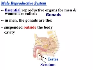
Male Reproductive System. Reproductive System. Primary sex organs (gonads): testes and ovaries Produce sex cells (gametes) Secrete steroid sex hormones Androgens (males) Estrogens and progesterone (females) Accessory reproductive organs: ducts, glands, and external genitalia.

E N D
Reproductive System • Primary sex organs (gonads): testes and ovaries • Produce sex cells (gametes) • Secrete steroid sex hormones • Androgens (males) • Estrogens and progesterone (females) • Accessory reproductive organs: ducts, glands, and external genitalia
Reproductive System • Sex hormones play roles in • Development and function of the reproductive organs • Sexual behavior and drives • Growth and development of many other organs and tissues
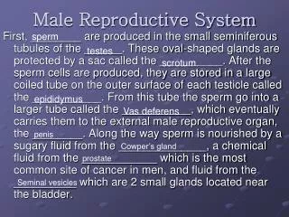
Male Reproductive System • Testes (within the scrotum) produce sperm • Sperm are delivered to the exterior through a system of ducts • Epididymis, ductus deferens, ejaculatory duct, and the urethra • Accessory sex glands: seminal vesicles, prostate, and bulbourethral glands • Empty secretions into the ducts during ejaculation
Ureter Urinary bladder Prostatic urethra Peritoneum Membranous urethra Seminal vesicle Ampulla of ductus deferens Urogenital diaphragm Pubis Ejaculatory duct Corpus cavernosum Corpus spongiosum Rectum Prostate Spongy urethra Bulbourethral gland Epididymis Glans penis Anus Prepuce Bulb of penis Testis Ductus (vas) deferens External urethral orifice Scrotum Figure 27.1
The Scrotum • Sac of skin and superficial fascia • Hangs outside the abdominopelvic cavity • Contains paired testes • 3C lower than core body temperature (temperature necessary for sperm production) • Temperature is kept constant by two sets of muscles • Smooth muscle that wrinkles scrotal skin (dartos muscle) • Bands of skeletal muscle that elevate the testes (cremaster muscles)
Urinary bladder Superficial inguinal ring (end of inguinal canal) Testicular artery Ductus (vas) deferens Spermatic cord Penis Autonomic nerve fibers Middle septum of scrotum Pampiniform venous plexus Cremaster muscle Epididymis External spermatic fascia Tunica vaginalis (from peritoneum) Superficial fascia containing dartos muscle Tunica albuginea of testis Scrotum Internal spermatic fascia Skin Figure 27.2
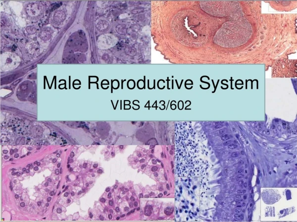
The Testes • Surrounded by two tunics • Tunica vaginalis, derived from peritoneum • Tunica albuginea, the fibrous capsule • Divided into 250–300 lobules, each containing 1–4 seminiferous tubules (site of sperm production) • Sperm are conveyed through • Seminiferous tubules • Tubulus rectus • Rete testis • Efferent ductules • Epididymis • Blood supply comes from the testicular arteries and testicular veins • Spermatic cord encloses nerve fibers, blood vessels, and lymphatics that supply the testes • Interstitial (Leydig) cells outside the seminiferous tubules produce androgens Testosterone
Spermatic cord Blood vessels and nerves Ductus (vas) deferens Testis Head of epididymis Seminiferous tubule Efferent ductule Rete testis Lobule Straight tubule Septum Tunica albuginea Body of epididymis Tunica vaginalis Duct of epididymis Cavity of tunica vaginalis Tail of epididymis (a) Figure 27.3a
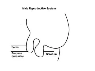
The Penis • External genitalia are the scrotum and the penis • Penis consists of • Root and shaft that ends in the glans penis • Prepuce, or foreskin—the cuff of loose skin covering the glans • Circumcision is the surgical removal of the foreskin • Crura: anchors penis to the pubic arch • Spongy urethra and three cylindrical bodies of erectile tissue (spongy network of connective tissue and smooth muscle with vascular spaces) • Corpus spongiosum surrounds the urethra and expands to form the glans and bulb • Corpora cavernosa are paired dorsal erectile bodies • Erection: erectile tissue fills with blood, causing the penis to enlarge and become rigid
Ureter Ampulla of ductus deferens Seminal vesicle Urinary bladder Ejaculatory duct Prostate Prostatic urethra Bulbourethral gland and duct Orifices of prostatic ducts Membranous urethra Urogenital diaphragm Bulb of penis Root of penis Crus of penis Bulbourethral duct opening Ductus deferens Corpora cavernosa Epididymis Corpus spongiosum Shaft (body) of penis Testis Section of (b) Spongy urethra Glans penis Prepuce (foreskin) (a) External urethral orifice Dorsal vessels and nerves Corpora cavernosa Urethra Skin Tunica albuginea of erectile bodies Deep arteries Corpus spongiosum (b) Figure 27.4
The Male Duct System • Epididymis • Ductus deferens • Ejaculatory duct • Urethra
Epididymis • Head: contains the efferent ductules • Duct of the epididymis • Microvilli (stereocilia) absorb testicular fluid and pass nutrients to stored sperm • Nonmotile sperm enter, pass slowly through, and become motile • During ejaculation the epididymis contracts, expelling sperm into the ductus deferens
Ductus Deferens and Ejaculatory Duct • Ductus deferens / Vas deferens • Passes through the inguinal canal • Expands to form the ampulla and then joins the duct of the seminal vesicle to form the ejaculatory duct • Propels sperm from the epididymis to the urethra • Vasectomy: cutting and ligating the ductus deferens, which is a nearly 100% effective form of birth control
Urethra • Conveys both urine and semen (at different times) • Has three regions • Prostatic urethra • Membranous urethra • Spongy (penile) urethra
Ureter Ampulla of ductus deferens Seminal vesicle Urinary bladder Ejaculatory duct Prostate Prostatic urethra Bulbourethral gland and duct Orifices of prostatic ducts Membranous urethra Urogenital diaphragm Bulb of penis Root of penis Crus of penis Bulbourethral duct opening Ductus deferens Corpora cavernosa Epididymis Corpus spongiosum Shaft (body) of penis Testis Section of (b) Spongy urethra Glans penis Prepuce (foreskin) (a) External urethral orifice Dorsal vessels and nerves Corpora cavernosa Urethra Skin Tunica albuginea of erectile bodies Deep arteries Corpus spongiosum (b) Figure 27.4
Accessory Glands: Seminal Vesicles • Produces viscous/thick alkaline seminal fluid • 70% of the volume of semen • Duct of seminal vesicle joins the ductus deferens to form the ejaculatory duct
Accessory Glands: Prostate • Encircles part of the urethra inferior to the bladder • Secretes milky, slightly acid fluid: • Plays a role in the activation of sperm • Enters the prostatic urethra during ejaculation
Accessory Glands: Bulbourethral Glands (Cowper’s Glands) • Pea-sized glands inferior to the prostate • Prior to ejaculation, produce thick, clear mucus • Lubricates the glans penis • Neutralizes traces of acidic urine in the urethra
Ureter Ampulla of ductus deferens Seminal vesicle Urinary bladder Ejaculatory duct Prostate Prostatic urethra Bulbourethral gland and duct Orifices of prostatic ducts Membranous urethra Urogenital diaphragm Bulb of penis Root of penis Crus of penis Bulbourethral duct opening Ductus deferens Corpora cavernosa Epididymis Corpus spongiosum Shaft (body) of penis Testis Section of (b) Spongy urethra Glans penis Prepuce (foreskin) (a) External urethral orifice Dorsal vessels and nerves Corpora cavernosa Urethra Skin Tunica albuginea of erectile bodies Deep arteries Corpus spongiosum (b) Figure 27.4
Semen • Mixture of sperm and accessory gland secretions • Contains nutrients (fructose), protects and activates sperm, and facilitates their movement (e.g., relaxin) • Prostaglandins in semen • Decrease the viscosity of mucus in the cervix • Stimulate reverse peristalsis in the uterus
Semen • Alkalinity neutralizes the acid in the male urethra and female vagina • Antibiotic chemicals destroy certain bacteria • Only 2–5 ml of semen are ejaculated, containing 20–150 million sperm/ml
Male Sexual Response • Erection • Enlargement and stiffening of the penis from engorgement of erectile tissue with blood • Initiated by sexual stimuli, including: • Touch, erotic sights, sounds, and smells • Can be induced or inhibited by emotions or higher mental activity • Parasympathetic reflex promotes release of nitric oxide (NO) • causes erectile tissue to fill with blood • Expansion of the corpora cavernosa • Compresses drainage veins and maintains engorgement • Corpus spongiosum keeps the urethra open • Impotence: the inability to attain erection
Male Sexual Response • Ejaculation • Propulsion of semen from the male duct system • Sympathetic spinal reflex causes • Ducts and accessory glands to contract and empty their contents • Bladder sphincter muscle to constrict, preventing the expulsion of urine • Bulbospongiosus muscles to undergo a rapid series of contractions
Spermatogenesis • Sequence of events that produces sperm in the seminiferous tubules of the testes • Most body cells are diploid (2n) and contain • Two sets of chromosomes (one maternal, one paternal) • 23 pairs of homologous chromosomes • Gametes are haploid (n) and contain • 23 chromosomes
Spermatogenesis • Spermatic cells give rise to sperm • Mitosis • Spermatogonia form spermatocytes • Meiosis • Spermatocytes form spermatids • Spermiogenesis • Spermatids become sperm
Cytoplasm of adjacent sustentacular cells Spermatogonium (stem cell) Sustentacular cell nucleus Basal lamina Type A daughter cell remains at basal lamina as a stem cell Type B daughter cell Tight junction between sustentacular cells Primary spermatocyte Secondary spermatocytes Early spermatids Late spermatids Cytoplasmic bridge Lumen of seminifer- ous tubule Spermatozoa (c) A portion of the seminiferous tublule wall, showing the spermato- genic cells surrounded by sustentacular cells (colored gold) Figure 27.7c
Mitosis of Spermatogonia • Begins at puberty • Spermatogonia • Stem cells in contact with the epithelial basal lamina • Each mitotic division a type A daughter cell and a type B daughter cell • Type A cells maintain the germ cell line at the basal lamina • Type B cells move toward the lumen and develop into primary spermatocytes
Basal lamina Type A daughter cell remains at basal lamina as a stem cell Spermatogonium (stem cell) Mitosis Type B daughter cell Growth Enters meiosis I and moves to adluminal compartment Primary spermatocyte Meiosis I completed Secondary spermatocytes Meiosis II Early spermatids Late spermatids Spermatozoa (b) Events of spermatogenesis, showing the relative position of various spermatogenic cells Figure 27.7b
Sperm • Major regions • Head: genetic region; nucleus and helmetlike acrosome containing hydrolytic enzymes that enable the sperm to penetrate an egg • Midpiece: metabolic region; mitochondria • Tail: locomotor region; flagellum
Approximately 24 days Golgi apparatus Acrosomal vesicle Mitochondria Acrosome Nucleus 1 2 Centrioles Spermatid nucleus Midpiece Head Microtubules 3 (a) Flagellum Excess cytoplasm 4 Tail 5 7 6 (b) Figure 27.8a, b
Hormonal Regulation of Male Reproductive Function • A sequence of hormonal regulatory events involving the hypothalamus, anterior pituitary gland, and the testes • Hypothalamus releases gonadotropin-releasing hormone (GnRH) • GnRH stimulates the anterior pituitary to secrete FSH and LH • FSH causes sustentacular cells to release androgen-binding protein (ABP), which makes spermatogenic cell receptive to testosterone • LH/ISCH stimulates interstitial cells to release testosterone • Testosterone is the final trigger for spermatogenesis • Feedback inhibition on the hypothalamus and pituitary results from • Rising levels of testosterone • Inhibin (released when sperm count is high)
1 GnRH Anterior pituitary Via portal blood 8 7 2 Inhibin 2 LH FSH Interstitial cells 4 3 6 Testosterone Somatic and psychological effects at other body sites Sustentacular cell Spermatogenic cells 5 Seminiferous tubule Stimulates Inhibits Figure 27.9
Male Secondary Sex Characteristics • Features induced in the nonreproductive organs by male sex hormones (mainly testosterone) • Appearance of pubic, axillary, and facial hair • Enhanced growth of the chest and deepening of the voice • Skin thickens and becomes oily • Bones grow and increase in density • Skeletal muscles increase in size and mass