Download
1 / 16
170 likes | 238 Vues
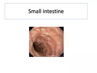
Small intestine II. jejunum and ileum. Dr. Anna L. Kiss. Department of Anatomy, Histology and Embryology Semmelweis University Budapest 2018. Jejunum, ileum. Small intestine: jejunum and ileum. jejunum+ileum. ascending colon. Peritoneal relationship of the small intestine.

E N D
Smallintestine II. jejunum and ileum Dr. Anna L. Kiss Department of Anatomy, Histology and Embryology Semmelweis University Budapest 2018
Small intestine: jejunum and ileum jejunum+ileum ascending colon
Peritoneal relationship of the small intestine • Radix mesenteri: oblique line • left: duodeno-jejunalflexure • right: sacroiliacjoint
Blood supply of the small intestine • Sup. mesenteric art. (fromtheabdominal aorta): • aa. jejunales et ilei
Blood supply of the small intestine Vasa recta Vasa recta
Histology of thesmallintestine T. mucosa: • epithelium: simple columnar (goblet cells) • propria (lymphoreticular connective tissue): glands (Lieberkhün crypts) • muscularis mucosae (2 layered smooth muscle) Submucosa: loose connective tissue; submucosus (Meissner) plexus; glands, lymphatic follicles) External muscle layer (t. muscularis): smooth muscle: inner circular, outer longitudinal) myenteric (Auerbach) plexus): intermuscular connective tissue Serosa or adventitia
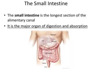
Histology of thesmallintestine • main part of the digestion and absorption • Histologically: accomodation to the function: increase the surface: Kerkring folds: plicae circulares (submucosa) villi: mucosa microvilli: apical plasma membrane of the enterocytes
Jejunum muscularis mucosae epithelium of mucosa intestinal villi Kerkrings’s folds mucosa submucosa tnica muscularis + serosa
Jejunum (longitudinal section of a villus) Goblet cells 1.) epithelium Mucosa layer stroma of villus 2.) propria: wide, containes Lieberkühn crypts: glands 3.) muscularis mucosae Lieberkühn’s crypts Submucosa
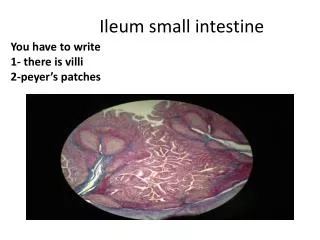
Ileum villi Peyer’s plaques crypts (Aggregated lymphatic follicles) submucosa muscle layer serosa
GALT About 70% of the body's immune systemis found in the digestive tract. The GALT is made up of several types of lymphoid tissue that produce and store immune cells that carry out attacks and defend against pathogens. Lymphoid tissue in the gut is comprised of the following : Tonsils and Adenoids (Waldeyer's ring) Peyer's patches in the small intestine Lymphoid aggregates in the appendix and large intestine Lymphoid tissue accumulating with age in the stomach Small lymphoid aggregates in the oesophagus Diffusely distributed lymphoid cells and plasma cells in the lamina propria of the gut
Bibliography • Lippert H: Lehrbuch Anatomie, Urban & Fischer, München, 2000 • Mac Kinnon P, Morris J: Oxford Lehrbuch der klinischen Anatomie, Hans Huber, Bern, 1997 • Snell RS, Clinical Anatomy, Little, Brown & Co, Boston, 1995 • Moore KL, Dalley AF: Clinically Oriented Anatomy, Lippincott, 1999