Download
1 / 98
980 likes | 984 Vues
UNIT 5. Medical Imaging. CT Scan. CT Scan. Computed axial tomography (CAT or CT scanning) is a medical imaging procedure that utilizes computer-processed X-rays to produce tomographic images or 'slices' of specific areas of the body.

E N D
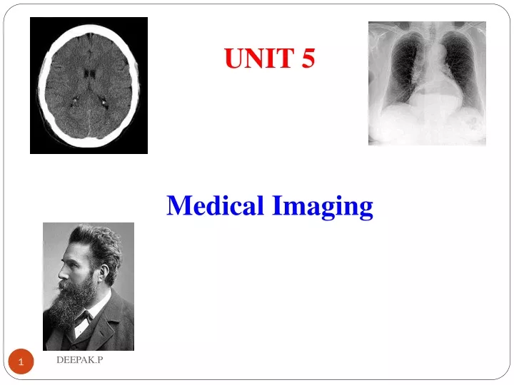
UNIT 5 Medical Imaging DEEPAK.P
CT Scan DEEPAK.P
CT Scan • Computed axial tomography (CAT or CT scanning) is a medical imaging procedure that utilizes computer-processed X-rays to produce tomographic images or 'slices' of specific areas of the body. • These cross-sectional images are used for diagnostic and therapeutic purposes in various medical disciplines. • Unlike other medical imaging techniques, such as conventional x-ray imaging (radiography), CT enables direct imaging and differentiation of soft tissue structures, such as liver, lung tissue, and fat. DEEPAK.P
CT Scan • Due to the short scan times of 500 milliseconds to a few seconds, CT can be used for all anatomic regions. DEEPAK.P
CT Scan Machine DEEPAK.P
CT Scan Principle • CT is based on the fundamental principle that the density of the tissue passed by the X-ray beam can be measured from the calculation of the attenuation coefficient. • The CT process involves several steps: • Scanning • Reconstruction • Visualization • Data collection DEEPAK.P
CT Scan Principle DEEPAK.P
CT Scan Principle DEEPAK.P
CT Scan Principle DEEPAK.P
Advantages of CT Scan • Relatively inexpensive compared with MRI and PET scanning • Accurate, 3-dimensional data including attenuation information • Rapid acquisition of data and no need for patients to remain for planning process DEEPAK.P
Components in CT Scan • 4 BASIC STEPS OF CT SCANNING • X-Ray Production • Data Acquisition • Data Processing • Image Display DEEPAK.P
Components in CT Scan DEEPAK.P
Components in CT Scan DEEPAK.P
Types of CT Scan • X-ray Computed Tomography: • This is the conventional CT scanner that scans a particular section of the subject at a time and sends it across to a computer for further processing • Spiral or Helical Computed Tomography: • These provide accurate information on the internal organs. • In spiral CT, the X-ray beam is emitted on a continuous basis and rotates around the subject, as the subject is moved through. • Micro Computed Tomography: In micro CT, the pixel size of the images is in micrometer. It is used in cases involving small animals, biomedical samples and other studies where minute detailing is desired. DEEPAK.P
Types of CT Scan • Micro Computed Tomography: • In micro CT, the pixel size of the images is in micrometer. • It is used in cases involving small animals, biomedical samples and other studies where minute detailing is desired. • Cone Beam Computed Tomography: • The Cone Beam CT is a recent addition, where the X-ray source is cone shaped and the resultant image is in 3-D. DEEPAK.P
Types of CT Scan DEEPAK.P
X- Ray DEEPAK.P
X-RAY • In 1895 Conrad Rontgen, a German physicist, discovered a previously unknown type of radiation while experimenting with gas-discharge tubes. • X-radiation (composed of X-rays) is a form of electromagnetic radiation. • X-rays have a wavelength in the range of 0.01 to 10 nanometers, • X-radiation is also called Rontgen radiation • An X-ray generator is a device used to generate X-rays. • The heart of an X-ray generator is the X-ray tube. DEEPAK.P
X-RAY DEEPAK.P
Electro Magnetic Spectrum DEEPAK.P
Uses of X- Ray DEEPAK.P
Uses of X-RAY • These devices are commonly used by radiographers to acquire an x-ray image of the inside of an object (as in medicine or non-destructive testing) but they are also used in sterilization or fluorescence. • X-ray machines are used in health care for visualizing bone structures and other dense tissues such as tumors. • Non-medicial applications include security and material analysis. • The two main fields in which x-ray machines are used in medicineare radiography and fluoroscopy. DEEPAK.P
Uses of X-RAY • Diagnostic still picture X-Ray • Examine bones and internal organs • Diagnostic continuous picture X-Ray(Fluoroscopy) • Examine internal organs as they functioning • Diagnostic motion picture X-Ray • Examine circulatory systems and its functioning • Diagnostic still picture X-Ray Scans • CT scan • Therapeutic X-Ray • For treatment DEEPAK.P
X-RAY in Radiography • Some forms of radiography include: • Orthopantomogram — a panoramic x-ray of the jaw showing all the teeth • Mammography — X-rays of breast tissue • Tomography — X-ray imaging in sections • Radiotherapy — the use of x-ray radiation to treat malignant cancer cells, a non-imaging application DEEPAK.P
X-RAY in Fluoroscopy • Fluoroscopy is used in cases where real-time visualization is necessary (and is most commonly encountered in everyday life at airport security). • Some medical applications of fluoroscopy include: • Angiography — used to examine blood vessels in real time • Barium enema — a procedure used to examine problems of the colon and lower gastrointestinal area • Barium swallow — similar to a barium enema, but used to examine the upper gastroinstestional area • biopsy — the removal of tissue for examination DEEPAK.P
X- Ray Tube DEEPAK.P
X-RAY Generation • Like any vacuum tube, the X-ray tube contains a cathode, which directs a stream of electrons into a vacuum, and an anode,whichcollects the electrons. • The anode in an X-ray tube is made of tungsten, molybdenum, or copper. • The electrons are then focused and accelerated by an electrical field towards an angled anode target. • The point where the electron beam strikes the target is called the focal spot. DEEPAK.P
X-RAY Generation • When electrons collide with the anode, about 1% of the resulting energy is emitted as X-rays, with the remaining 99% released as heat. • A cooling system is necessary to cool the anode; many X-ray generators use water or oil re-circulating systems • The intensity of X rays depends on the current through the tube. • This current can be varied by varying the heater current. DEEPAK.P
X-RAY Generation • The wavelength of the X rays depends on the target material and the velocity of the electrons hitting the target. • It can be varied by varying the target voltage of the tube. DEEPAK.P
X ray tube DEEPAK.P
Generation of X- ray from X ray tube DEEPAK.P
Block Diagram DEEPAK.P
X-RAY Generation • The intensity of X rays depends on the current through the tube. • This current can be varied by varying the heater current. • The wavelength of the X rays depends on the target material and the velocity of the electrons hitting the target. • It can be varied by varying the target voltage of the tube. DEEPAK.P
Simple Block Diagram DEEPAK.P
Block Diagram DEEPAK.P
Collimators DEEPAK.P
Collimators • A collimator is a device that narrows a beam of particles or waves. • In optics, a collimator may consist of a curved mirror or lens with some type of light source and/or an image at its focus. DEEPAK.P
Collimators in X-rays • In X-ray, and gamma ray optics, a collimator is a device that filters a stream of rays so that only those traveling parallel to a specified direction are allowed through. DEEPAK.P
Collimators in X-rays DEEPAK.P
Collimators DEEPAK.P
Collimators in X-rays • This may be a sheet of lead or other material opaque to the incoming radiation with many tiny holes bored through it. • Only rays that are travelling nearly parallel to the holes will pass through them—anyothers will be absorbed by hitting the plate surface or the side of a hole. • It allows the radiographer to control the exposure of radiation to expose a film. DEEPAK.P
Gantry detectors DEEPAK.P
Gantry detectors • The gantry is the 'donut' shaped part of the CT scanner that houses the components necessary to produce and detect x-rays to create a CT image. • The x-ray tube and detectors are positioned opposite each other and rotate around the gantry aperture. • This gantry can rotate 360 degrees around its axis. DEEPAK.P
Gantry detectors DEEPAK.P
Gantry detectors • gantry aperture (720mm diameter) • microphone • sagittal laser alignment light • patient guide lights • x-ray exposure indicator light • emergency stop buttons • gantry control panels • external laser alignment lights • patient couch • ECG gating monitor DEEPAK.P
Gantry detectors • CT scanner with cover removed to show internal components. T: X-ray tubeD: X-ray detectorsX: X-ray beamR: Gantry rotation DEEPAK.P
X-Ray Detectors DEEPAK.P
X-Ray detectors • The two most common types of X-ray detector used are the scintillation and the gas-filled detectors. • Scintillation detectors work by converting x-rays to optical photons in special materials and then detecting the light with a photomultiplier tube or a photodiode. • The X-ray photon collides with a phosphor screen, or scintillator and produces photons in the blue region of the visible spectrum. • These are subsequently converted to voltage pulses by means of a photomultiplier tube attached directly behind the scintillator. DEEPAK.P
X-Ray detectors • A gas filled detector consists of a rectangular gas cell with thin entrance and exit windows. • Inside the detector, an electric field of about 100 V/cm is applied across two parallel plates. • Some of the x-rays in the beam interact with the chamber gas to produce fast photoelectrons, DEEPAK.P
Solid State X-ray Detectors • X-ray interacts in material to produce photoelectrons which are collected by applying a drift field DEEPAK.P