Download
1 / 69
700 likes | 708 Vues
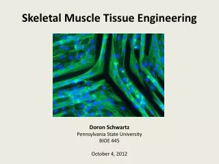
Skeletal Muscle 骨骼肌. Qiang XIA ( 夏强 ), PhD Department of Physiology Zhejiang University School of Medicine. Tel: 88206417, 88208252 Email: xiaqiang@zju.edu.cn. Skeletal muscle 骨骼肌. Cardiac muscle 心肌. Smooth muscle 平滑肌. Skeletal muscles are attached to the skeleton by tendons.

E N D
Skeletal Muscle 骨骼肌 Qiang XIA (夏强), PhD Department of Physiology Zhejiang University School of Medicine Tel: 88206417, 88208252 Email: xiaqiang@zju.edu.cn
Skeletal muscle骨骼肌 • Cardiac muscle 心肌 • Smooth muscle平滑肌
Skeletal muscles are attached to the skeleton by tendons. Skeletal muscles typically contain many, many muscle fibers.
The sarcomere(肌小节) is composed of: thick filaments called myosin, anchored in place by titin fibers, and thin filaments called actin, anchored to Z-lines .
A cross section through a sarcomere shows that: • each myosin can interact with 6 actin filaments, and • each actin can interact with 3 myosin filaments.
Myosin filament (thick filament)粗肌丝 • Myosin肌凝蛋白
Actin filament (thin filament)细肌丝 • Actin肌纤蛋白 • Tropomyosin原肌凝蛋白 • Troponin肌钙蛋白
Sarcotubular system (1) Transverse Tubule横管 (2) Longitudinal Tubule纵管 Sarcoplasmic reticulum肌浆网
Contraction (shortening): myosin binds to actin, and slides it, pulling the Z-lines closer together, and reducing the width of the I-bands. Note that filament lengths have not changed.
Contraction: myosin’s cross-bridges(横桥) bind to actin; the crossbridges then flex to slide actin.
The thick filament called myosin is actually a polymer of myosin molecules, each of which has a flexible cross-bridge that binds ATP and actin.
4. Partial hydrolysis of the bound ATP energizes or “re-cocks” the bridge. 2. The full hydrolysis and departure of ADP + Picauses the flexing of the bound cross-bridge. 3. Binding of a “new” ATP to the cross-bridge uncouples the bridge. The myosin-binding site on actin becomes available, so the energized cross-bridge binds. 1. The cross-bridge cycle requires ATP
The myosin-binding site on actin becomes available, so the energized cross-bridge binds. 1.
2. The full hydrolysis and departure of ADP + Picauses the flexing of the bound cross-bridge.
3. Binding of a “new” ATP to the cross-bridge uncouples the bridge.
4. Partial hydrolysis of the bound ATP energizes or “re-cocks” the bridge.
4. Partial hydrolysis of the bound ATP energizes or “re-cocks” the bridge. 2. The full hydrolysis and departure of ADP + Picauses the flexing of the bound cross-bridge. 3. Binding of a “new” ATP to the cross-bridge uncouples the bridge. The myosin-binding site on actin becomes available, so the energized cross-bridge binds. 1. The cross-bridge cycle requires ATP
Roles of troponin, tropomyosin, and calcium in contraction In relaxed skeletal muscle, tropomyosin blocks the cross-bridge binding site on actin. Contraction occurs when calcium ions bind to troponin; this complex then pulls tropomyosin away from the cross-bridge binding site.
Excitation-contraction coupling 兴奋-收缩偶联 • Transmission of action potential (AP) along T tubules • Calcium release caused by T tubule AP • Contraction initiated by calcium ions
The latent period between excitation and development of tension in a skeletal muscle includes the time needed to release Ca++ from sarcoplasmic reticulum, move tropomyosin, and cycle the cross-bridges.
The transverse tubules bring action potentials into the interior of the skeletal muscle fibers, so that the wave of depolarization passes close to the sarcoplasmic reticulum, stimulating the release of calcium ions. The extensive meshwork of sarcoplasmic reticulum assures that when it releases calcium ions they can readily diffuse to all of the troponin sites.
Passage of an action potential along the transverse tubule opens nearby voltage-gated calcium channels, the “ryanodine receptor,” located on the sarcoplasmic reticulum, and calcium ions released into the cytosol bind to troponin. The calcium-troponin complex “pulls” tropomyosin off the myosin-binding site of actin, thus allowing the binding of the cross-bridge, followed by its flexing to slide the actin filament.
Which of these following proteins contains the binding sites for Ca2+ that initiates contraction? A Myosin B Troponin I C Tropomyosin D Troponin C E Troponin T
General process of excitation and contraction in skeletal muscle 骨骼肌兴奋与收缩的基本过程 • Neuromuscular transmission • Excitation-contraction coupling • Muscle contraction
A single motor unit(运动单位) consists of a motor neuron and all of the muscle fibers it controls.
1. The exocytosis of acetylcholine from the axon terminal occurs when the acetylcholine vesicles merge into the membrane covering the terminal. 2. On the membrane of the muscle fiber, the receptors for acetylcholine respond to its binding by increasing Na+ entry into the fiber, causing a graded depolarization. 3. The graded depolarization typically exceeds threshold for the nearby voltage-gate Na+ and K+ channels, so an action potential occurs on the muscle fiber.
Nicotinic acetylcholine receptor 烟碱型乙酰胆碱受体 Acetylcholinesterase 乙酰胆碱酯酶
Miniature end plate potential 微终板电位 • Small fluctuations (typically 0.5 mV) in the resting potential of postsynaptic cells. • They are the same shape as, but much smaller than, the end plate potentials caused by stimulation of the presynaptic cell. Miniature end plate potentials are considered as evidence for the quantal release of neurotransmitters at chemical synapses, a single miniature end plate potential resulting from the release of the contents of a single synaptic vesicle.
A woman comes to your clinic and explains that she is noting gradually worsening fatigue/weakness in her legs when she goes for her walk. She also mentions a droopy right eyelid, and wonders if this is a normal aging process. You examine her and find the following: overall decreased muscle strength, trace reflexes throughout, and weakness of eyelid closure bilaterally. The rest of the exam is unremarkable. What would you administer to treat the likely condition? A Muscarinic blockers B Nicotinic blockers C Acetylcholinesterase blockers D Alpha blockers E Beta blockers
A woman comes to your clinic and explains that she is noting gradually worsening fatigue/weakness in her legs when she goes for her walk. She also mentions a droopy right eyelid, and wonders if this is a normal aging process. You examine her and find the following: overall decreased muscle strength, trace reflexes throughout, and weakness of eyelid closure bilaterally. The rest of the exam is unremarkable. What would you administer to treat the likely condition? A Muscarinic blockers B Nicotinic blockers C Acetylcholinesterase blockers D Alpha blockers E Beta blockers
Neuromuscular transmission A Is caused by the release of acetylcholine from the muscle side of the junction B Shows a permeability change to Na+ and K+ at the receptor site during the endplate potential (EPP) C May be facilitated by curare in myasthenia gravis D Is blocked by curare because it competes with the Na+ entry during the muscle action potential E Is solely an electronic function
Neuromuscular transmission A Is caused by the release of acetylcholine from the muscle side of the junction B Shows a permeability change to Na+ and K+ at the receptor site during the endplate potential (EPP) C May be facilitated by curare in myasthenia gravis D Is blocked by curare because it competes with the Na+ entry during the muscle action potential E Is solely an electronic function
A miniature end-plate potential is A Not related to changes in ionic permeability B A reduced action potential in the motor end-plate C Produced by spontaneous release of acetylcholine D Responsible for weak muscular contractions E An afterdischarge at the neuromuscular junction
A miniature end-plate potential is A Not related to changes in ionic permeability B A reduced action potential in the motor end-plate C Produced by spontaneous release of acetylcholine D Responsible for weak muscular contractions E An afterdischarge at the neuromuscular junction
The action of acetylcholine at the neuromuscular junction is terminated primarily by A Enzymatic breakdown by choline acetylase B Enzymatic breakdown by acetylcholinesterase C Uptake into the muscle D Uptake into the nerve ending E Diffusion into the surrounding extracellular fluid
The action of acetylcholine at the neuromuscular junction is terminated primarily by A Enzymatic breakdown by choline acetylase B Enzymatic breakdown by acetylcholinesterase C Uptake into the muscle D Uptake into the nerve ending E Diffusion into the surrounding extracellular fluid
The transmission of an action potential over the muscle fiber membrane causes the contraction of the fiber a few milliseconds later. Which of the following terms is used to describe that process? A Ratchet Theory of Muscle Contraction B Excitation Contraction Coupling C Membrane Potential D All-or -Nothing Law
The transmission of an action potential over the muscle fiber membrane causes the contraction of the fiber a few milliseconds later. Which of the following terms is used to describe that process? A Ratchet Theory of Muscle Contraction B Excitation Contraction Coupling C Membrane Potential D All-or -Nothing Law
Mechanics of single-fiber contraction • Muscle tension 肌张力 – the force exerted on an object by a contracting muscle • Load 负荷 – the force exerted on the muscle by an object (usually its weight) • Isometric contraction 等长收缩 – a muscle develops tension but does not shorten (or lengthen) (constant length) • Isotonic contraction 等张收缩 – the muscle shortens while the load on the muscle remains constant (constant tension)
Twitch contraction 单收缩 • The mechanical response of a single muscle fiber to a single action potential is know as a TWITCH
iso = same tonic = tension metric = length Tension increases rapidly and dissipates slowly Shortening occurs slowly, only after taking up elastic tension; the relaxing muscle quickly returns to its resting length.
All three are isotonic contractions. • Latent period潜伏期 • Velocity of shortening • Duration of the twitch • Distance shortened