Download
1 / 12
160 likes | 208 Vues
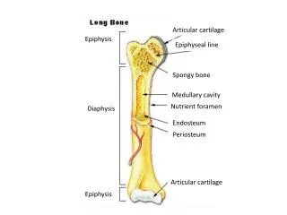
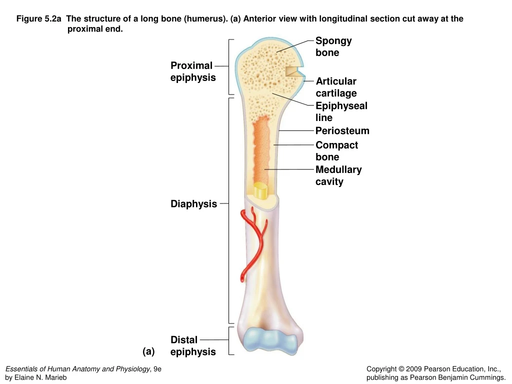
Figure 5.2a The structure of a long bone (humerus). (a) Anterior view with longitudinal section cut away at the proximal end. Spongy bone. Proximal epiphysis. Articular cartilage. Epiphyseal line. Periosteum. Compact bone. Medullary cavity. Diaphysis. Distal epiphysis. (a).

E N D
Figure 5.2a The structure of a long bone (humerus). (a) Anterior view with longitudinal section cut away at the proximal end. Spongy bone Proximal epiphysis Articular cartilage Epiphyseal line Periosteum Compact bone Medullary cavity Diaphysis Distal epiphysis (a)
Figure 5.2a The structure of a long bone (humerus). (a) Anterior view with longitudinal section cut away at the proximal end.
Figure 5.3a Microscopic structure of compact bone. (a). Osteon (Haversian system) Lamellae Blood vessel continues into medullary cavity containing marrow Spongy bone Perforating fibers Compact bone Periosteal blood vessel Central (Haversian) canal Periosteum Perforating (Volkmann’s) canal (a) Blood vessel
Figure 5.10 Paranasal sinuses. Sphenoid sinus Frontal sinus Frontal sinus Ethmoid sinus Ethmoid sinus Maxillary sinus Sphenoid sinus Maxillary sinus (a) (b)
Figure 5.10 Paranasal sinuses. (a) (b)
Figure 5.29 General structure of a synovial joint. Acromion of scapula Joint cavity containing synovial fluid Ligament Bursa Ligament Articular (hyaline) cartilage Tendon sheath Synovial membrane Fibrous articular capsule Tendon of biceps muscle Humerus
Figure 5.30 Types of synovial joints. (f) Carpals Nonaxial Uniaxial (b) Biaxial Multiaxial (c) Humerus Ulna (a) Plane joint Ulna Radius (a) (b) Hinge joint (e) (d) (c) Pivot joint Metacarpal Phalanx Carpal Metacarpal #1 (d) Condyloid joint Head of humerus Scapula (e) Saddle joint (f) Ball-and-socket joint
Figure 5.30 Types of synovial joints. (f) Carpals Nonaxial Uniaxial (b) Biaxial Multiaxial (c) Humerus Ulna Ulna Radius (a) (e) (d) Metacarpal Phalanx Carpal Metacarpal #1 Head of humerus Scapula