Download
1 / 36
370 likes | 385 Vues
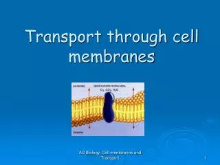
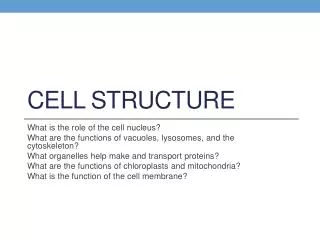
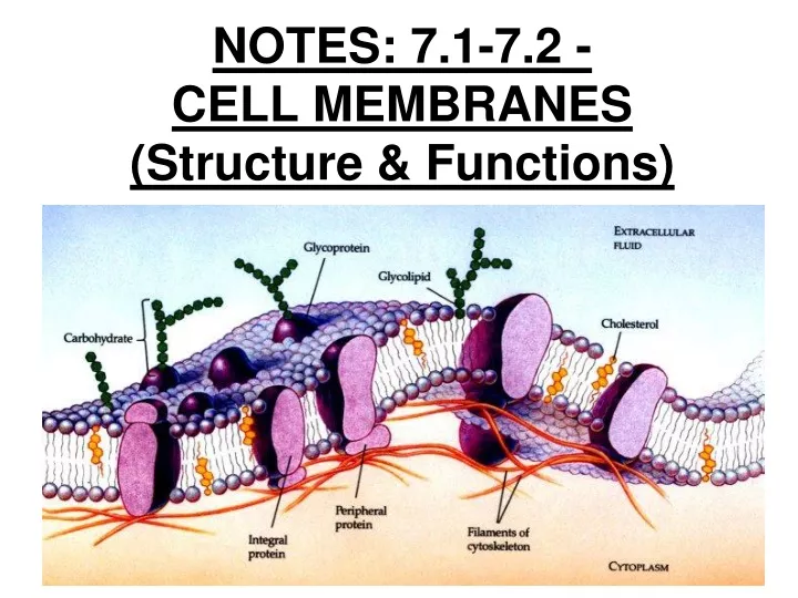
NOTES: 7.1-7.2 - CELL MEMBRANES (Structure & Functions). Membranes = “the edge of life”. Overview: Life at the Edge. ● The plasma membrane is the boundary that separates the living cell from its nonliving surroundings

E N D
Overview: Life at the Edge ● The plasma membrane is the boundary that separates the living cell from its nonliving surroundings ● The plasma membrane exhibits selective permeability, allowing some substances to cross it more easily than others
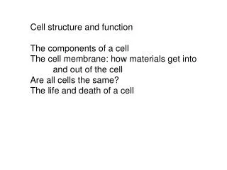
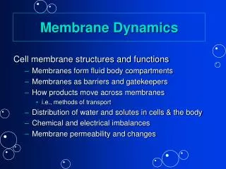
What must a membrane do? ● let some things leave cell / enter cell ● keep stuff inside or outside of cell ● display cell antigens / markers ● hold receptors for big molecules ● be able to seal, fuse, be flexible as cell changes shape
When we understand the cell membrane, we will better understand: ● diseases like CF, diabetes ● how to better design drug delivery systems ● transplants without dangerous immunosuppressive drugs ● how to better design anesthetics
CELL MEMBRANE STRUCTURE: ● Imagine if you coated a cell with an impermeable shell...life would cease! ● UNLESS...you allowed for doors and windows in the shell! **This is what living cells do...every cell is encased within a lipid membrane which contains doors and windows made of PROTEINS.
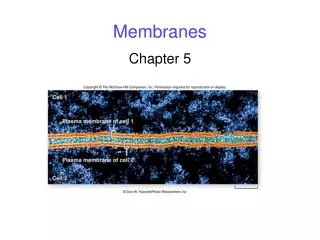
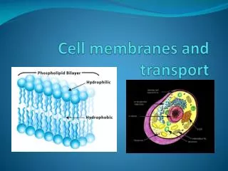
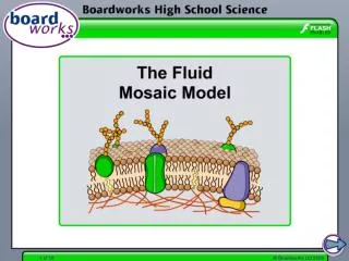
7.1 - Cell membranes are fluid mosaics of lipids and proteins ● Phospholipids are the most abundant lipid in the plasma membrane ● Phospholipids are amphipathic molecules, containing hydrophobic and hydrophilic regions ● The fluid mosaic model states that a membrane is a fluid structure with a “mosaic” of various proteins embedded in it
● The lipid layer that forms the foundation of cell membranes is composed of molecules called PHOSPHOLIPIDS.
● one end is extremely polar (hydrophilic); ● one end is strongly nonpolar (hydrophobic)
WATER Hydrophilic head Hydrophobic tail WATER
● the cell membrane forms spontaneously in water because the nonpolar lipid “tails” are repelled by polar water molecules; the polar “heads” of the molecules form hydrogen bonds with water molecules.
● So, every phospholipid molecule orients so that its polar “head” faces water and its nonpolar “tails” face away... two layers are formed with the tails facing each other...the result is called a LIPID BILAYER.
● the interior of the bilayer is completely nonpolar; therefore it repels any water-soluble (polar or ionic) molecules that try to pass through. ● (So, if a cell were fully encased within a pure lipid bilayer, it would be completely impermeable to water-soluble molecules like sugars, polar amino acids, proteins, salts, etc.)
7.2 - Lipid bilayer membranes are: ●PERMEABLE to: -lipids -nonpolar molecules: O2, CO2 -small polar molecules: H2O ●IMPERMEABLE to: -ions (Na+, K+, Cl-) -large polar molecules: sugars, proteins
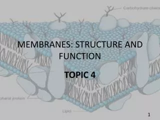
Membrane Proteins and Their Functions (7.1 & 7.2): ● A membrane is a collage of different proteins embedded in the fluid matrix of the lipid bilayer ● Proteins determine most of the membrane’s specific functions ●Peripheral proteins are not embedded ●Integral proteins penetrate the hydrophobic core and often span the membrane
Fibers of extracellular matrix (ECM) Glycoprotein Carbohydrate Glycolipid EXTRACELLULAR SIDE OF MEMBRANE Cholesterol Peripheral proteins Microfilaments of cytoskeleton Integral protein CYTOPLASMIC SIDE OF MEMBRANE
●Integral proteins that span the membrane are called transmembrane proteins ● The hydrophobic regions of an integral protein consist of one or more stretches of nonpolar amino acids, often coiled into alpha helices
Hydrophilic region of protein Phospholipid bilayer Hydrophobic region of protein
EXTRACELLULAR SIDE N-terminus C-terminus CYTOPLASMIC SIDE a Helix
Functions of Membrane Proteins: ● Six major functions of membrane proteins: -Transport -Enzymatic activity -Signal transduction -Cell-cell recognition -Intercellular joining -Attachment to the cytoskeleton and extracellular matrix (ECM)
Signal Enzymes Receptor ATP Enzymatic activity Transport Signal transduction
Glyco- protein Attachment to the cytoskeleton and extra- cellular matrix (ECM) Cell-cell recognition Intercellular joining
3 Main Types of Membrane PROTEINS… 1) Channels / Transport 2) Receptor proteins 3) Cell surface markers
1. CHANNELS / TRANSPORT: ● a given channel will transport only certain kinds of molecules...which gives the cell membrane its selectively permeable nature
Transport Proteins (7.2): ● Transport proteins allow passage of hydrophilic substances across the membrane ● Some transport proteins, calledchannel proteins, have a hydrophilic channel that certain molecules or ions can use as a tunnel ● Channel proteins called AQUAPORINSfacilitate the passage of water
● Other transport proteins, called carrier proteins, bind to molecules and change shape to shuttle them across the membrane ●A transport protein is specific for the substance it moves!
2. RECEPTOR PROTEINS: ● collect & transmit information from the cell’s environment; a receptor protein will bind a molecule on the outer surface of the cell (i.e. a hormone) which causes a change on the other end of the protein on the inner surface of the cell; this triggers a response within the cell **LIGAND: molecule that binds to specific receptor site on another molecule
3. CELL SURFACE MARKERS: ● many proteins (or lipids in some cases) have carbohydrate “flags” (usually <15 monomers) attached to the exterior end of the protein; identify your body‘s cells as belonging to YOU -useful in cell-cell recognition (e.g. sorting of animal embryo’s cells into tissues and organs; rejection of foreign cells by the immune system)
“GLYCO” = carbohydrate, so: *glycoprotein = protein w/ carbo chain *glycolipid = lipid w/ carbo chain
The Role of Membrane Carbohydrates in Cell-Cell Recognition ● Cells recognize each other by binding to surface molecules, often carbohydrates, on the plasma membrane ● Membrane carbohydrates may be covalently bonded to lipids (forming glycolipids) or more commonly to proteins (forming glycoproteins) ● Carbohydrates on the external side of the plasma membrane vary among species, individuals, and even cell types in an individual
CHOLESTEROL: ● acts as a “fluidity buffer” for the membrane ● resists changes in fluidity that can occur with changes in temp. ● found in cell membranes of eukaryotes; makes the membrane: 1) less fluid at warmer temps. 2) more fluid at lower temps.