Download
1 / 106
1.15k likes | 1.4k Vues
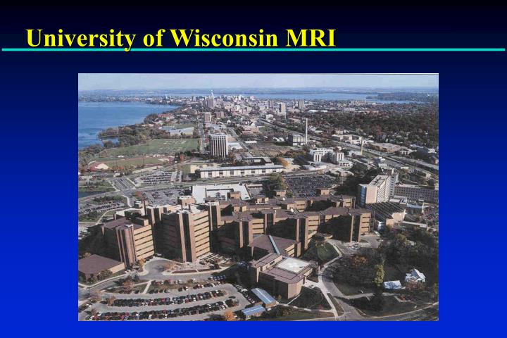
University of Wisconsin MRI. Magnetic Resonance Angiography. Walter Block, Ph.D. Charles Mistretta, Ph.D. Thomas Grist, MD Departments of Biomedical Enginering, Radiology and Medical Physics University of Wisconsin - Madison. Collaborators. Faculty: Chuck Mistretta, Ph.D.

E N D
Magnetic Resonance Angiography Walter Block, Ph.D. Charles Mistretta, Ph.D. Thomas Grist, MD Departments of Biomedical Enginering, Radiology and Medical Physics University of Wisconsin - Madison
Collaborators Faculty: Chuck Mistretta, Ph.D. Tom Grist, M.D. Kris Pillai, M.D. Funding: Whitaker Foundation NIH HBL Institute Students and Graduates: Dana Peters, Ph.D. Andrew Barger, Ph.D. Oliver Wieben, Ph.D. Ethan Brodsky Tian Liang Gu Aiming Lu
MRA Methods Time-of-Flight Phase Contrast Contrast-Enhanced
ky kx k-Space Signal Image
ky kx k-Space Signal Image Detail Contrast
5 % 10 % 20 % 50 %
Objectives • Describe three acquisitiontechniques • Time of Flight (TOF) • Contrast-enhanced MRA (CE-MRA) • Phase Contrast (PC) • 4D MRA ( time-resolved 3D MRA) • Discuss post-processing methods throughout
Objectives • Describe three acquisition techniques • Time of Flight (TOF) • Contrast-enhanced MRA (CE-MRA) • Phase Contrast (PC) • 4D MRA ( time-resolved 3D MRA) • Discuss post-processing methods throughout
RF Saturation Initial Value 1.0 0.5 0 RF Pulses Longitudinal Magnetization (along B0) Regrowth Equilibrium magnetization TR Time
Flow Related Enhancement Imaging Slice Flow 0 1 2 Mz Static Tissue Time
Flow Related Enhancement Vessels Static Tissue Image from Anderson, Edelman and Turski Clinical MRA
2D Sequential TOF Acquisition slice advance
MIP Images from 2D TOF Volume Data coronal axial source image F. Korosec PhD Thesis U. of Wisconsin 1991 sagittal right carotid sagittal left carotid
Saturation Pulses Saturation Volume Imaging Slice
Saturation Pulses Isolate Arteries and Veins superior sat (arteries) inferior sat (veins) no sat F. Korosec, PhD Thesis, U. of Wisconsin, 1991
2D TOF Acquisition Scan Time(TOF) = TR • NEX • # PE • # slices Advantages • Good inflow contrast • Trailing sat used for venous suppression • Retrospective reprojection Disadvantages • Voxel size greater in slice direction • Greater intravoxel dephasing due to large voxel and longer TE due to large slice select gradient amplitude • In-plane vessels can saturate • Pulsatility artifacts possible in ungated scans • Susceptible to motion
Pulsatility Artifact Unsaturated spins Saturated spins Systole Diastole Reduce flip angle to reduce saturation
Pulsatility Artifact tip angle = 90 tip angle = 30 Anderson. Edelman, and Turski, Clinical MRA
= SNR Voxel size * ADC time 3D TOF Acquisition
3D Acquisition Scan time(TOF) = TR • NEX• # PE • # SE Advantages • More isotropic spatial resolution • Reduced intravoxel dephasing • Shorter TE than 2D • High SNR • Retrospective reprojection Disadvantages • Potential saturation in distal portions of volume • Saturation of slow flow
Objectives • Describe three acquisition techniques • Time of Flight (TOF) • Contrast-enhanced MRA (CE-MRA) • Phase Contrast (PC) • 4D MRA ( time-resolved 3D MRA) • Discuss post-processing methods throughout
Theory: T1 Shortening of Gadolinium • During rapid IV infusion, Gadolinium concentrated in arteries for 1 min. • Gadolinium is a potent T1 relaxation agent in blood • T1blood 1200 ms <100 ms • Arterial MR signal enhancement is proportional to T1 shortening
0 60 Effect of Gadolinium on MRA Signal Through Plane 0 Gado 60 T1 = 50 Signal In-Plane 0 15 T1 = 1000 Number of RF Pulses
Pre-contrast Slice Post-contrast Slice
2D TOF vs. 3D Contrast MRA 2D TOF 3D CE-MRA
Why use CE-MRA? • Fast: scans less than 1 minute • TR drops from 40 ms to 5ms • Free to orient slab along vessels, instead of perpendicular • easy to perform • eliminates respiratory motion artifact • Short TE • less sensitive to signal loss at a stenosis • less sensitive to susceptibility artifact • Uniform blood signal • independent of blood flow velocity • no “in-plane flow” saturation artifact
Renal CE-MRA Renal vein Renalartery Signal Renal parenchyma 0 10 20 30 40 Time (s)
Contrast preparation • MR compatible power injector • Gadolinium not nephrotoxic • Excreted by glomerular filtration • Load 0.3 mmol/kg Gd-DTPA (up to 40 cc) • Load 50 cc saline flush
Scan time for CE-MRA Scan time = TR x (Phase Enc) x (Slice Enc) • TR = repetition time • Phase Enc = # phase encoding steps • Slice Enc = number of slices Select parameters for scan <35 sec.
Methods to reduce image acquisition time • Shorter TR • Faster gradients • More efficient pulse sequences • Higher bandwidth • Partial Sampling of k-space • Partial Fourier (Echo, Readout, Phase encoding, Slice encoding), Projection Undersampling
TR=4.7 TR=8.5 RBW=64kHz RBW=32kHz SNR=22.9 SNR=44.0
Methods to reduce image acquisition time • Shorter TR • Faster gradients • More efficient pulse sequences • Higher bandwidth • Partial Sampling of k-space • Partial Fourier (Echo, Readout, Phase encoding, Slice encoding), Projection Undersampling
ky kx k-Space Signal Image
ky ky kx kx k-Space Signal kx • Partial Fourier • Fractional Echo
Zero Interpolation Processing ZIP x2 & ZIP 512 No ZIP
ZIP 512 No ZIP
Prescribing Volume Image Volume Monitor Volume
Timing • Injection begins prior to 3D CE-MRA • Variable scan delay • Patient instructed to hold breath • 3D CE-MRA acquisition begins
Arterial Venous Tissue Timing of MRA scan relative to bolus
Timing Artifacts Poorly timed Properly timed Maki, Prince et al JMRI 1996, 6:642-651
Arteries & Veins Arteries
Coordination of Contrast Arrival Timing Scan • 2 cc test bolus • One sagittal slice per second Smart Prep • Large monitor voxel • Automatic trigger Fluoro Triggering • Real-time system • Thick 2D image @ 1 frame/s • Custom hardware Hany, JMRI 1997 Foo, Radiology 1997 Riederer, MRM 1997