Download
1 / 36
370 likes | 575 Vues
Integumentary System. Consists of the skin, plus all the appendages (or accessory structures) of the skin including : Sweat glands Oil Glands Hair Nails. General Functions of the Integumentary System. Protection from injury Protect from pathogens Sensation Thermoregulation.

E N D
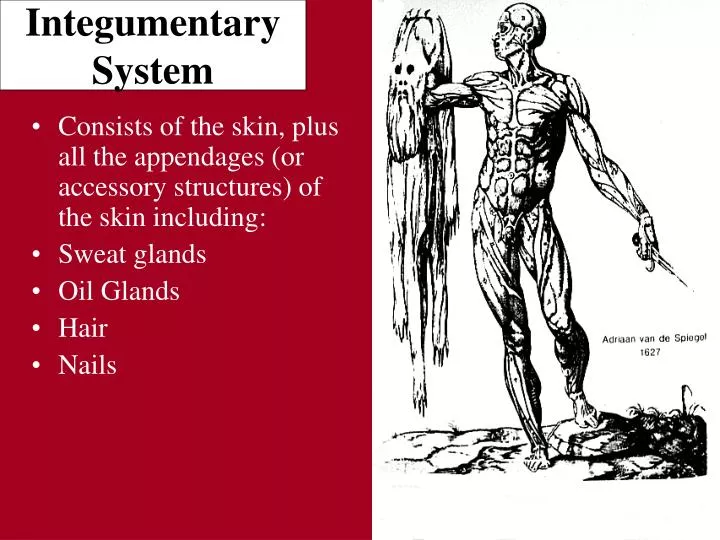
Integumentary System • Consists of the skin, plus all the appendages (or accessory structures) of the skin including: • Sweat glands • Oil Glands • Hair • Nails
General Functions of the Integumentary System • Protection from injury • Protect from pathogens • Sensation • Thermoregulation
Basic Skin Structure • The skin has 2 main layers: • The superficial epidermis -4-5 layers of epithelial cells resting upon a basement membrane. • The deep dermis consisting of fibrous connective tissue. Contains blood vessels, oil glands, sweat glands
Identify the epidermis and the dermis! Which is made of connective tissue? What type?
The Epidermis • Epithelial Tissue. • Consists of 4 distinct cell types arranged in 4 or 5 distinct layers. Yellow arrow indicates the epidermis of thick skin
Skin Types • Thick Skin • Found on soles of feet and palms of hands • Contains 5 epidermal layers or strata (“sheets”): (from base up) • Stratum basale • Stratum spinosum • Stratum granulosum • Stratum lucidum • Stratum corneum
Skin Types • Thin skin • Found everywhere else. • Contains only 4 layers. (lacks a stratum lucidum). • Why is thick skin found on the palms and soles? What is the advantage of that? Note: this slide is at a higher mag. than the thick skin slide on the previous page
Notice the 4 layers of thin skin in both the cartoon and the photomicrograph.
Skin Color • Due to 3 pigments: • Melanin • Carotene • Hemoglobin • Of these, only melanin is made in the skin. • Melanin: • Albinos lack thispigment. • Ranges in color from yellow to reddish brown to black. • Freckles and moles are local accumulations of melanin.
Carotene • Yellow to orange pigment found in plant products such as carrots. • Hemoglobin • Pigmented protein that transports oxygen within the blood. • Inlight skinned people, the fair skin allows the crimson color of oxygenated blood to make the skin have a somewhat pinkish hue.
Dermis • Strong, flexible fibrous connective tissue. • Provides an arena for immune cells to fight invaders. • Heavily invested with blood vessels • Contains multiple sensory receptors. • Collagen fibers give the skin strength. • Elastingives the skin the ability to stretch and recoil.
Hypodermis • A layer of adipose (fat) below the dermis • Contains blood vessels en route to the dermis and the roots of hair follicles.
Appendages of the Skin • Oil glands • Sweat glands • Hair • Nails 1 4 2
Oil Glands • Simple glands found everywhere except palms of the hands and soles of the feet. • Secrete an oily, lipid-rich secretion called sebum. • Sebum softens and lubricates the skin. It also decreases the skin’s permeability to water and is quite bactericidal.
The oil gland is indicated by the arrow. Note how its duct empties into a hair follicle.
Sweat Glands • Distributed over the entire body except the nipples and portions of the external genitalia. • Over 2.5 million per person.
Hair and Hair Follicles • Hair is a flexible strand made of dead cells. • Large amounts of keratin in both hair and nails. • The hair is made by the living hair follicle.
Hair and Hair Follicles • Hair consists of a shaft which protrudes from the skin and the root which is within the skin. At the base, the root gets wider forming the hair bulb.
Hair and Hair Follicles • The hair follicle surrounds much of the hair root. • The hair papillacontains the blood vessels that nourish the matrix and the cells of the hair follicle.
Notice the hair shaft, hair follicle, papilla, and the multiple oil glands.
Hair and Hair Follicles • Wrapped around the bulb of the follicle is a network of sensory nerve endings known as the hair root plexus. Allows the hairs to serve a sensory function. • Attached to each hair is a bundle of smooth muscle known as an arrectorpili muscle. In times of fright or cold, these muscles contract and cause the hair to stand on end – and produces goose bumps.
The arrow indicates an arrector pili muscle. In this picture, you should also try to identify the shaft, root, follicle, hair papilla, and sebaceous gland.
Skin Cancer • Because of its role as our external covering, the skin takes a tremendous amount of abuse. • One serious disorder that can result is skin cancer. • Cancer can be thought of as uncontrolled cell division and growth. • There are 3 types of skin cancers we will discuss: • Basal cell carcinoma • Squamous cell carcinoma • Malignant melanoma From abnormal cells, a cancerous cell develops Cancerous cells spread, forming a tumor An abnormal cell develops
Skin Cancer • Basal cell carcinoma • Most common (70% of skin cancers) • Least vicious • Usually cured via surgical removal • Affects stratum basale.
Skin Cancer • Squamous cell carcinoma • Arises from stratum spinosum. • 25% of cases. • Good prognosis if caught and treated early • Can be fatal if it metastasizes to the lymph nodes.
Skin Cancer • Malignant melanoma • Least common and most dangerous. • Often arises from a pre-existing mole. • Follow the ABCD rule for early detection: Asymmetry (2 sides do not match) Border irregularity Color (multiple) Diameter (>6mm is bad!)
First Degree Burns • Only the epidermis is damaged • Heals in 2-3 days • i.e. Sunburn
Second Degree Burns • Injury to epidermis and upper region of dermis • Red and painful, blisters appear • No scarring if infection is avoided
Third Degree Burns • Destroy all layers of the skin • Burned area appears white, gray, or blackened • Nerve endings are destroyed, so burn is not painful.
Other infections and allergies: • Athlete’s Foot- fungus resulting in itchy, red, peeling condition • Cold Sores- Herpes simplex virus, when active causes small, fluid filled blisters usually around the lips and mouth. • Impetigo- water-filled, raised lesions that rupture caused by a highly contagious staph infection. • Psoriasis- chronic condition. Overproduction of skin cells resulting in reddened lesions.