Download
1 / 63
990 likes | 2.09k Vues
PULMONARY PATHOLOGY. LECTURE 6 RESPIRATORY SYSTEM DEPARTMENT OF HEALTH SCIENCES DR SHAI WEEK OF NOVEMBER 10, 2013. OBJECTIVES. Upper Respiratory Tract Disorders Inflammatory conditions Neoplasms Lung Diseases COPD Emphysema Asthma Bronchitis Neoplasms Interstitial diseases.

E N D
PULMONARY PATHOLOGY LECTURE 6 RESPIRATORY SYSTEM DEPARTMENT OF HEALTH SCIENCES DR SHAI WEEK OF NOVEMBER 10, 2013
OBJECTIVES • Upper Respiratory Tract Disorders • Inflammatory conditions • Neoplasms • Lung Diseases • COPD • Emphysema • Asthma • Bronchitis • Neoplasms • Interstitial diseases
Inflammatory Conditions Upper Respiratory Tract • Rhinitis: • Inflammation of nasal mucosa • Acute or chronic • Acute • Aetiology: infection (viral: rhinovirus, respiratory syncytial virus, influenza, corona) or allergy (hay fever) type I hypersensitivity reaction • Signs: blocks nasal airways • Chronic infection / allergy causing chronic inflammation • Signs: Macroscopic: nasal polyps, microscopic: oedematous tissue with inflammatory cells, eosinophils
Sinusitis • Inflammation of the sinuses • Acute • Acute maxillary sinusitis (ethmoid & frontal less common) • Aetiology: 2ndary to rhinitis • Pathogenesis: inflammation in sinus lining, mucosa swells> stasis of maxillary sinus secretions> 2ndary bacterial infection> purulent mucous • Chronic • Chronically thick & inflammed mucosa of sinus • Aetiology: acute sinusitis, cigarette smoke, industrial exposure, polyps
Necrotizing Lesions • A. Mucormycotic lesions • Opportunistic* fungal infections of nose from immunosuppression> fatal unless treated rapidly • B. Lymphoma • Progressive ulceration & destruction of structures in nose, sinus, form of T cell lymphoma • Histologically: infiltration of lymphocytes, plasma cells, blast cells, lymphoid cells, fatal if untreated
Neoplasms • 1. Nasopharyngeal angiofibroma • Males, age 10 – 25 yo • Mimics malignant tumour during puberty • Ulceration & bleeding common • 2. Plasmacytomas • Malignant tumour • Composed of monoclonal plasma cells* • Presents as soft, haemorrhagic nasal mass • 3. Olfactory neuroblastoma • Rare, upper nasal cavity • Haemorrhagic mass, bone destruction, metastases in 20% cases
4. Nasopharyngeal Carcinoma • Squamous or anaplastic carcinoma of nasopharynx • Abundant lymphoid tissue in stroma • Associated with Epstein-Barr virus • Small, undetected until lymph node swelling metastases in neck • Prognosis: good with radiation therapy • 5 year survial: 80% if localized, 50% if advanced
The larynx – inflammatory conditions • A. Acute Laryngitis • Acute inflammation of larynx, infective, allergic or irritative (eg smoking) • Sequelae • Resolution: without complications • Spread: tracheobronchitis develops • Airway obstruction: layngealoedemaegHaemophilus influenza • B. Croup • Acute inflammation & obstruction of RESP TRACT (larynx, trachea, bronchi) • Children 6 months to 3 years
Croup aetiology: viral with 2ndary bacterial infection • Signs of obstruction: stridor (difficulty breathing), tachycardia, cyanosis, restlessness • Management: humidification, intubation or tracheostomy*
Reactive Nodules • Polyps: common benign lesions associated with URTI (upper respiratory tract infections) after vocal abuse> polypectomy • Singer’s Nodules: smooth, round nodules at junction between anterior 1/3 and posterior 2/3 of vocal cords • Singers, oedematous connective tissue with submucosal fibrosis covered by squamous epithelium
papillomas • Papilloma: warty papillomas on the larynx due to infection with Human Papillomavirus (HPV 11, 16), Solitary lesions • Juvenile Papillomatosis: multiple soft, pink papillomas on vocal cords in children • Histologically: florid viral warts, persistent & recurrent, requires multiple excisions
Sqaumous cell carcinoma of larynx • 2% of cancers, incidence 2 per 100,000 per year worldwide • > 40 years age, males more than females • Risk factors: smoking, radiation • Affected sites: • supraglottic region (30%) eg epiglottis • Glottic region (60%) true vocal cords, anterior and posterior commissures • Subglottic region (10%): below the true vocal cords, above the 1st tracheal ring • Macroscopically: ulcerated, gray papillary lesions • Microscopically: Well differentiated, keratinizing squamous carcinomas • Spread: local, lymph, haematogenous
Disorders of the lungs • Atelectasis • Defective expansion & collapse of lungs • As a result of: • Obstruction {of large bronchial tubes leads to resorption of air from lung distal to obstruction- inhaled foreign bodies, bronchial cancer, TB, large lymph nodes, lung cancer} • Compression {compression of lung by accumulation of fluid or air in pleural cavity} • Scarring {causes contraction of parenchyma} • Surfactant loss {failure of lung expansion} • Outcomes: expansion may be aided by physiotherapy and bronchoscopy mediated removal
Chronic obstructive pulmonary disease • Obstructive vs restrictive lung diseases • Obstructive: obstruction to air flow within lungs, although lungs may be hyper inflated. If chronic > COPD chronic obstructive pulmonary disease • Restrictive: obstruction to EXPANSION of lungs (fibrosis or oedema) so that they can only take in a limited amount of air • Lungs are under inflated, rate of airflow unaffected • Eg: pulmonary fibrosis • Both obstructive & restrictive lung disease: impairs pulmonary function
Mechanisms: by which airflow may be reduced in COPD causing 2 clinical pictures • 1) increased airway resistance, by narrow airways eg bronchitis, bronchiectasis, asthma results in hypercapnea* hypoxemia*, cyanosis • 2) decreased outflow pressure due to loss of elastic recoil of lungs, eg emphysema • Diagnosis & treatment: CHEST X RAY • * SEE hyperinflation, flat hemi diaphragms, reduced peripheral vascular markings, bullae • And lung function tests • Physiotherapy • Bronchodilators • Antibiotics • Note: respiratory centers of some patients with COPD are insensitive to CO2 , relying on hypoxic drive to maintain respiration. Therefore, it is dangerous to give 02 without careful observation
emphysema • Permanent dilatation of any part of the air spaces DISTAL to terminal bronchiole with or without destruction of tissue, but with NO SCARRING • Common conditions, increasing with age, males more than females • Aetiology: includes smoking, atmospheric pollution, family history • Associated with chronic bronchitis, and alpha 1 anti trypsin deficiency • Pathogenesis: extra cellular proteases secreted into lung by inflammatory cells are inhibited by protease inhibitors (alpha 1 anti trypsin) • Theses inhibitors are inactive (by smoke) or absent, resulting in continued activity of proteases with destruction of lung parenchyma • Leads to loss of elastic recoil in lungs and decreased area of available gaseous exchange.
Types of emphysema • Defined by location of damage in respiratory acinus • 1. Centrilobar: dilatation of respiratory bronchioles at centre of acinus, lesions in upper lobes • 2. pan lobar: dilatation of terminal alveoli, later affects bronchioles and whole acinus, affects lower lobes • 3. paraseptal: air spaces at periphery of loules, adjacent to pleura • 4. irregular: scarring, trapped air following lung fibrosis
Clinical features • Early stage: rapid respiratory rate enables individuals to maintain blood oxygenation, so levels of C02 and 02 are near normal, “breathless pink puffers” not cyanoses, but on exertion, hypoxic • Later stage: reduced O2 uptake even at rest. Devline in respiratory function, cyanosis, hypercapnea, corpulmonale (right heart failure) • CXR: • Hyperinflated, trachea descended (decreased distance
Chronic bronchitis • Cough productive of sputum on most days for 3 months of the year for at least 2 successive years • Middle aged men, associated with smoking • Pathogenesis: • Irritation by cigarette smoke causes inflammation of the respiratory bronchioles (bronchiolitis) and increased mucus secretion • Hyper secretion of mucus associated with hypertrophy, and hyperplasia of bronchiole mucus secreting glands • The Reid index: gives the ratio of gland to wall thickness in the bronchus, • It is significantly increased in chronic bronchitis • Bronchiolar obstruction must be extensive and widespread to give symptoms • Clinical features: early stages: chronic cough with sputum • Later stages: disease progresses to more severe, hypoxaemia, cyanosis, hypercapnea, corpulmonale, respiratory failure
Bronchial asthma • Increased irritability of bronchial tree, with narrowing of airways, may reverse spontaneously or after broncho dilator treatment. • Triggers • Allergy: allergans* trigger IgE mediated type I hypersensitivity (dust mites, food, animal danders, drugs) • Infection: respiratory tract infection can trigger bronchoconstriction • Occupational exposure: allergens, direct irritation • Drugs: beta antagonists, aspirin • Irritant gases: sulphur dioxide, nitric oxide, ozone smog • Psychological stress, cold air, exercise
Asthma is associated with atopic disease, eczema, hay fever, some allergies • Asthma can be classified as • 1) Extrinsic (atopic): early onset asthma, triggered by allergens, individuals have IgE levels raised, commonest type of asthma • 2) Intrinsic: non atopic: late onset asthma, triggered by infection of URT. IgE levels are normal, no family history,skin testing is negative • Pathogenesis: both types, obstruction is caused by a combination of bronchospasm, oedema, mucus plugging
Phases • 1: early (15 minutes): rapid onset of bronchoconstriction, caused by histamine release from mast cell degranulation, the allergen binds to IgE antibodies on surface of mast cells • 2. late (5 hours): second wave of bronchoconstriction, after initial recovery, inflammatory mediators released by mast cells cause activation of macrophages & chemotaxis of polymorphs & eosinophils into bronchial mucosa, these cells release inflammatory mediators causing 2nd wave of bronchoconstriction • 3. prolonged hyperreactivity (days): exaggerated response of airway on further re exposure to allergen. There is persistent inflammation leading to bronchial wall damage.
Structural changes in asthma • Immune cell infiltration: bronchial mucosa is infiltrated with eosinophils, mast cells, lymphoid cells, macrophages • Mucosa oedema & hypersecretion of mucus: plugs airways • Hypertrophy of bronchial smooth muscle due to recurrent bronchoconstriction • Focal necrosis of airway epithelium • Deposition of collagen in epithelium • Sputum contains Charcot-Leyden crystals (from eosinophil granules) & Curschmann’s spirals (mucus plugs from small airways)
Clinical features of asthma • Mild: intermittenet* episodes of bronchospams • Moderate: severe and irreversible asthma in middle age (chronic asthma) • Status asthmaticus: severe, acute distress, does not respond to drug therapy, air entry inadequate> silent chest is ominous sign*, death from respiratory insufficiency • Signs: wheeze, barrel chest • Complications: corpulmonale, pulmonary hypertension • Management: successfully managed with b2 adrenoreceptor agonists, corticosteroids, aminophylline, anticholinergics, cromoglycate
Bronchiectasis • Irreversible dilatation of bronchi • Congenital: cystic fibrosis, kartageners syndrome (bronchiectasis, dextrocardia, sinusitus) • Acquired: infection (whooping cough, pneumonia, measles) and obstruction (inhaled foreign body) • Haemophilus influenza and pseudomonaaeruginosa are commonest pathogens
Cystic fibrosis • Hereditary multisystem disease • Lack of production of abnormally thick mucus, primarily affecting lungs & pancreas • Commonest autosomal recessive disorder, 1/2000 newborns incidence. • 1/25 Caucasians* are heterozygous* carriers of CF gene
CF PATHOGENESIS • Mutated gene on chromosome 7, encodes for protein termed “cystic fibrosis transmembrane regulator” CFTR • Commonest mutation is deletion of phenylalanine residue at position 508 • Normaly this protein enables transport of chloride ions across cell membranes • In CF: a defective CFTR results in impaired chloride transport, which prevents furtehr release of sodium and water to liquefy mucus • Net result: production of EXTEREMELY THICK mucus by exocrine glands
Mucus obstructs: bronchi, intestine, pancreas • Leading to: congested lungs, meconiumileus in newborn bowel, malabsorption and failure to thrive • Respiratory issues: repeated infections (s. aureus, pseudomonas), bronchiectasis, hyper inflation of lungs from trapped air, hypoxia scarring and destruction of pulmonary vascular bed.
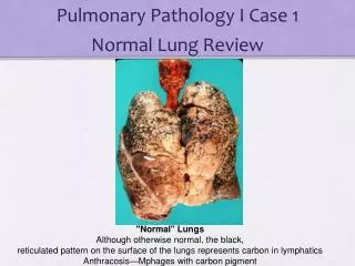
Infections of the lung • Pneumonia is defined as the consolidation of lung tissue caused by the formation of intra-alveolar inflammatory exudates, from a long standing lung infection • Risk factors • Suppressed cough in coma • Impaired mucociliary clearance (smoke, gases, viruses, immotile cilia) • Pulmonary oedema from Rt sided cardiac failure • Impaired alveolar macrophages (alcohol, smoke, 02 toxicity) • Immunosuppresion, drugs, instrumentation, disability, immobility
Classification of pneumonia • Bacterial pneumonia (80%) • Clinical: fever, short of breath, cough, sputum, coarse/crackles • Bronchopneumonia • Infection on bronchi, but inflammatory exudate into alveoli, patchy consolidation of lung • Affects young or hospital acquired • Pathogenesis: patients develop retention of secretions which gravitate to dependent parts of the lungs and becomes infected • Macroscopically: bilateral, multiple areas of consolidation, bronchial mucosa is inflamed and pus around peripheral bronchi • Microscopically: acute inflammation of bronchi, inflammatory exudate in lumina and alveoli
Outcomes of pneumonia • Resolves • Bronchial damage • Lung fibrosis • Lung abcess formation • Empyema (pus in pleural cavity) • Pericarditis • Death
Lobar pneumonia • Uniform consolidation of part of a lobe, from infection. • 20-50 years age, poor social conditions • Pneumococcus, klebsiella • Pathogenesis: • Organisms enter distal air spaces without colonization of bronchi • Infection spreads rapidly into alveolar spaces • Macroscopically: whole lobe becomes consolidated and airless • Microscopically: alveoli are filled with inflammatory exudate
Pulmonary tuberculosis • TB • Chronic granulomatous infection of lung from Mycobacterium Tuberculosis • Leading cause of death in parts of Africa and Asia • Affects older people, HIV infected, immigrant populations (Hajj issue) • Spread by: • Inhalation of M. tuberculosis droplets (commonest) • Ingestion of spoiled food or milk • Inoculation of skin* • Transplacental (congenital)
Pathogenesis of tb • DESTRUCTION FROM HYPERSENSITIVITY reaction of host directed against bacterial wall constituents • Sequence • 1. 0-10 days,mycobacteria start inflammatory response. Neutrophilsphagozytose organisms, but cannot destroy them, so engulfed bacteria are drained into lymph nodes • 2. after 10 days: development of T cell mediated immune response (type IV hypersensitivity), to bacillary* cell wall results in cytokine release, leading to activation of macrophages> chronic inflammatory pattern, dominated by epitheliod cells which form GRANULOMAS • Central core of necrotic caseous tissue containing viable mycobacteria • Tuberculousgranulomas are termed TUBERCLES
Morphology of tb • Macroscopically: granulomas are pin sized white/gray tubercles in tissues • Microscopically: granulaomas are the histological hallmark of TB • Granuloma: central area of caseous necrosis, surrounded by 3 layers • 1st layer: activated macrophages (Langhans giant cells) • Middle layer: lymphocytes • Outer layer: fibroblastic tissue • Healing of granuloma is slow, with progressive fibrosis and calcification (CXR) • The central necrotic area may remain caseous and REACTIVATION results in secondary infection
Primary and secondary tb • Primary TB: 1st encounter with organism, resulting in development of small parenchymal focus, large lymph drainage • Secondary infection: reactivation of previously infected indvidual, results in large localized parencyymal reaction, minimal lymph node involvement
Neoplastic diseases of lung • Bronchogenic Carcinoma • Commonest cause of death from neoplasm in UK • >30,000 cases per year • Males more, 40-70 years peak • Risk factors • Cigarette smoking (early start-increased risk), decline if smoking stops • Occupational: asbestos*, nickel, radioactive material • Environmental: radon • Pulmonary fibrosis • Histological types (4) • 1. squamous cell carcinoma (50%) • 2. small cell carcinoma (oat cell, 20%) • 3. adenocarcinoma (20%) • Large cell anaplastic carcinoma (10%)