Download
1 / 100
1.11k likes | 2.24k Vues
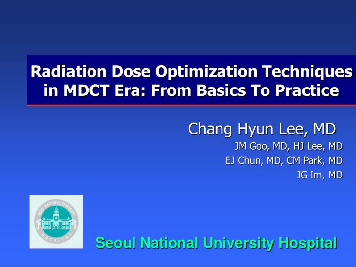
Radiation Dose Optimization Techniques in MDCT Era: From Basics To Practice. Chang Hyun Lee, MD JM Goo, MD, HJ Lee, MD EJ Chun, MD, CM Park, MD JG Im, MD. Seoul National University Hospital. Purpose. To discuss the importance of radiation dose modulation in MDCT

E N D
Radiation Dose Optimization Techniques in MDCT Era: From Basics To Practice Chang Hyun Lee, MD JM Goo, MD, HJ Lee, MD EJ Chun, MD, CM Park, MD JG Im, MD Seoul National University Hospital
Purpose • To discuss the importance of radiation dose modulation in MDCT • To review the characteristics of radiation dose modulation techniques in different MDCT scanners • To explain how to use and apply this techniques to the patients during MDCT scan
Contents • Basic concepts in CT dose index • Fundamental dose parameters • CT dose index • Parameters affecting CTDI • Radiation dose increase in MDCT scanner - Radiation dose in MDCT - Radiation risk • Effective dose in various CT examination • Effective dose • Calculation and estimates of effective dose • Radiation dose summary • Radiation dose modulation techniquesin different MDCT scanners –Reference mAs–Reference noise index– it’s advantages and disadvantages • Practical tipsfor optimizing radiation dose in MDCT
Fundamental Dose Parameters BACK TO CONTENTS
Fundamental Dose Parameters 10 mSv = 1 rem
Fundamental Dose Parameters • Exposure • Roentgens (C/kg) or air kerma (J/kg – mGy) • Ionization in air per unit mass or amount of energy imparted per unit mass • Related to intensity of radiation at point of measurement • Irradiated area, penetrating power, tissue sensitivity에 대한 고려가 없음.
Fundamental Dose Parameters • Absorbed dose • Joules/kg or mGray (mGy) • Energy absorbed by material per unit mass • Depends on radiation type, energy & material irradiated • Quoted locally or averaged over area e.g. organ • Absorbed dose– amount of radiation energy deposited in the patient’s body as a result of exposure • Radiation exposure: radiation source-related term • Radiation dose: body-related term D D ~ 3D
Fundamental Dose Parameters • Effective dose • mSv • Measure of radiation risk to patient • Attempts to reflect equivalent whole body dose that results in same stochastic risk • Applies organ sensitivity factors • Enables risk comparison between different procedures and modalities
Fundamental Dose Parameters NOTES • Absorbed dose, however, does not account for differing sensitivities of the organs to radiation damage. • Equivalent dose in a tissue is a product of the tissue type and the radiation weighting factor • Equivalent dose has the same numerical value as absorbed dose and is measured in sievert or rem. • Effective dose is computed by summing the absorbed doses in the organsweighted by their radiation sensitivity. • Effective dose estimates the whole-body dose that would be required to produce the same risk as partial-body dose delivered by a localized radiological procedure - useful in evaluating potential biological risk of a specific radiologic examination.
CT Dose Index BACK TO CONTENTS
CT dose index • CTDI was developed originally by Shope et al. back in 1981. • The basic radiation dose parameter in CT is the CT dose index (CTDI). • Represents absorbed radiation dosein aCT dose phantom, measured in the gray or rad.
CT dose index: CTDI • CTDI has been defined for use with single-detector row CT scanners. • CTDI is the total energy absorbed within a dose profile deposited within one nominal collimation.
CT dose index • Three derivatives of the CTDI • CTDI100: radiation exposure measured by means of an ionization chamber with a length of 100 mm. • CTDI100w: weighted radiation dose in axial (nonhelical) CT scan • CTDIvol: volume CTDI = CTDIw/pitch
CT dose index: CTDI (Multiple-scan average dose)
Measurement equipment: CTDI • Ionization chamber • Thermoluminescent Dosimeters (TLD) • X-ray film
Average dose in scan plane: CTDIw • Weighted average CTDI represents the average dose in scan plane of Perspex phantom CTDIw = [2/3 CTDI100 (periphery) + 1/3 CTDI100 (center)] x 33.7
Average dose in scanned volume: CTDIvol • Axial: CTDIvol=CTDIw x (slice width/couch inc.) • Helical: CTDIvol = CTDIw / Pitch Noncontiguous exposure along z-axis
CTDI • Advantages of CTDI as a dose descriptor • Disadvantages of CTDI
Parameters affecting CTDI BACK TO CONTENTS
Effect of scan parameters on CTDIvol • mA and scan time (mAs per rotation) • CTDIvol increase linearly with mA and scan time • E.g 2 x mAs = 2 x CTDIvol
Variation of CTDIvol with kVp • CTDIvol increases with kVp • Approx ∝kVp2 ImPact 2005
CTDI and slice width • CTDI increases if irradiated width does not match nominal width CTDI = Area / nT Z-axis T1 T2 T3
Variation of CTDIvol with no. of slices • Number of slices • CTDIvol is independent of number of slices • Absorbed dose: energy absorbed per unit mass
Effect of pitch on CTDIvol • CTDIvol is inversely proportional to pitch • E.g. doubling pitch halves the CTDIvol • … but only if mA remains constant • On some systems mA automatically adjusted for pitch so CTDIvol is constant
Effect of patient size • For same scan parameters (mAs, kV) the dose increases as phantom/patient size decreases • For pediatrics CTDI can underestimate dose by ~ x 2 if measured in standard sized phantoms
Radiation dose in MDCT BACK TO CONTENTS
Radiation dose in MDCT • ~ 60 million CTs per year – doubled in 5 years • > 60,000 CT examinations at University of Alabama at Birmingham, USA • ~ 67% total radiation exposure is from CT
Radiation Dose in MDCT Y Imanishi et al. Eur Radiol (2005) 15:41-46
CT examination is increasing in Japan No. of image (x10,000) No. of exam/year (x1,000) No. of CT Nishizawa, Acta Radiol Jap 2004; 64:151-158
Scan area is also increasing Nishizawa, Acta Radiol Jap 2004; 64:151-158
Does multi-slice CT impart more or less radiation dose? • An increase by 10-30% may occur with multi-slice detector array ICRP (International Commission on Radiological Protection) Publication 87
Doses from Chest CT Helical CT MDCT (4) LDCT (single) kV 120 120 120 mA 100-210 300-350 25 Rotation time (s/r) 1-1.5 0.5 2 Table feed (mm/s) 10 7-11 20/2s/r FOV (cm) 27 35 30 Organ dose (mGy) Bone marrow 5.91 7.19 2.51 Lung 20.94 19.59 3.09 Stomach 8.59 19.83 1.41 Breast 18.24 20.20 2.41 Liver 0.43 19.62 1.64 Esophagus 18.12 18.16 2.90 Thyroid 8.23 23.70 2.41 Effective dose (mSv) 7.6211.01.40 Nishizawa, Acta Radiol Jap 2004; 64:suppl
Radiation Risk BACK TO CONTENTS
Audience Response Question • What is the conventional estimate of the long term risk of death from cancer for 10 mSv whole body exposure for an average person? • A. Negligible • B. 1:20,000 • C. 1:2,000 • D. 1:200
Audience Response Question • What is the conventional estimate of the long term risk of death from cancer for 10 mSv whole body exposure for an average person? • A. Negligible • B. 1:20,000 • C. 1:2,000 • D. 1:200
Audience Response Question • The radiation dose from a chest radiograph is approximately what fraction of the dose from yearly natural background radiation? • A. 1:100 • B. 1:10 • C. 1:1 • D. 10:1
Audience Response Question • The radiation dose from a chest radiograph is approximately what fraction of the dose from yearly natural background radiation? • A. 1:100 • B. 1:10 • C. 1:1 • D. 10:1
Audience Response Question • The radiation dose for One CT scan versus One chest radiograph? • A. CT < CR • B. CT > CR, CT < 10 CR • C. CT > CR, 10 CR < CT < 100 CR • D. CT > CR, CT = 100 ~ 250 CR • E. CT > CR, CT > 500 CR
Audience Response Question • The radiation dose for One CT scan versus One chest radiograph? • A. CT < CR • B. CT > CR, CT < 10 CR • C. CT > CR, 10 CR < CT < 100 CR • D. CT > CR, CT = 100 ~ 250 CR • E. CT > CR, CT > 500 CR
Relative Dose Beliefs Diagnostic CT Scans: Assessment of Patient, Physician, and Radiologist Awareness of Radiation Dose and Possible Risks. Lee CL et al. Radiology 2004
Cancer Risk Beliefs Diagnostic CT Scans: Assessment of Patient, Physician, and Radiologist Awareness of Radiation Dose and Possible Risks. Lee CL et al. Radiology 2004
Radiation Cancer Risk Conventional Theory • 5% excess cancer deaths per Sv (100 rem) • 1:2,000 (0.05%) excess cancer deaths per 10 mSv (1 rem) Brenner et al. Estimated Radiation Risks Potentially Associated with Full-Body CT Screening. Radiology 2004
Age-Dependent Cancer Risk Brenner et al. Estimated Radiation Risks Potentially Associated with Full-Body CT Screening. Radiology 2004
Radiation Risk in Context • Baseline risk of cancer in life time 20-25% • Younger patients at higher risk • Late middle aged adult getting average CT has life time risk of cancer increased from ~20% to ~20.01% • Not measurable
Radiation – Common Doses and Risks • Chest radiograph (adult): 0.02 mSv • 0.0001 – 0.000002% deaths • UGI/BE: 1-7 mSv • 0.005-0.035% deaths • Mammography2.6 mGy, but • Effective Dose 0.13 mSv (so cancer risk much lower) • Natural background: 3 mSv/yr • Medical population average 0.3-0.6 mSv/yr
Comparable Non-Radiation Risks • Assume 10 mSv (1 rem) CT scan • Smoking 140 cigarettes in a lifetime (lung cancer) • Spending 7 months in New York City (air pollution – lung cancer) • Driving 4,000 miles in a car (accident) • Flying 250,000 miles in a jet (accident)
Audience Response Question • A single CT scan has a lifetime risk of death from cancer similar to: • Smoking 7 cigarettes • Driving 40,000 miles in a car • Having a standard IVU • Flying into low earth orbit