Download
1 / 48
630 likes | 1.37k Vues
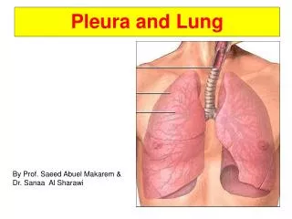
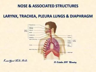
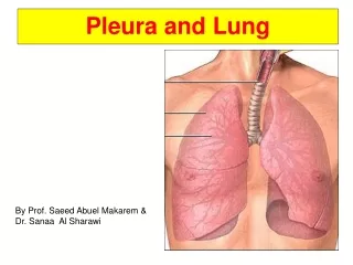
Pleura and Lungs . Pleura . The pleura is divided into two major types, based on location: 1. Parietal pleura 2. Visceral pleura Each pleural cavity is the potential space enclosed between the visceral and parietal pleurae. Parietal pleura . 1. Costal part 2. Diaphragmatic part

E N D
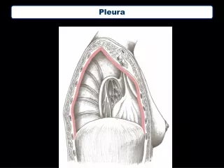
Pleura • The pleura is divided into two major types, based on location: • 1. Parietal pleura • 2. Visceral pleura • Each pleural cavity is the potential space enclosed between the visceral and parietal pleurae.
Parietal pleura • 1. Costal part • 2. Diaphragmatic part • 3. Mediastinal part • 4. Cervical part
Parietal pleura has 4 parts • Costal pleura-Lining internal surface of thoracic wall • Mediastinal pleura- Covering sides of mediastinum • Diaphragmatic pleura-Covering superior surface of dome of each hemidiaphragm • Cervical pleura-A dome of pleura extending superiorly into superior thoracic aperture
Visceral pleura • Covers the lungs • Cannot be dissected from lung • Insensitive to pain
Nerve Supply of the Pleura • The parietal pleura is sensitive to pain, temperature, touch, and pressure and is supplied as follows: • The costal pleura is segmentally supplied by the intercostal nerves. • The mediastinal pleura is supplied by the phrenic nerve. • The diaphragmatic pleura is supplied over the domes by the phrenic nerve and around the periphery intercostal nerves.
visceral pleura • The visceral pleura covering the lungs is sensitive to stretch but is insensitive to common sensations such as pain and touch. It receives an autonomic nerve supply from the pulmonary plexus .
Pleural recesses • 1. Costomediastinal recesses • 2. Costodiaphragmatic recesses
Lines of Pleural Reflection Costomediastinal recesses Costodiaphragmatic recesses
Pleural reflections MCL MAL Vertebral • Lungs : 6th rib 8th rib 10thvert • Pleura : 8th rib 10 th rib 12 thvert
Pleural effusion • Excess fluid that accumulates in pleural cavity • Can impair breathing by limiting the expansion of lungs during inhalation • Types • Serous fluid (hydrothorax) • Blood (hemothorax) • Chyle (chylothorax) • Pus (pyothorax or empyema)
Thoracocentesis • To obtain a sample of pleural fluid or to remove blood or pus or air • To avoid damage to intercostal nerve and vessels, needle is inserted superior to rib, high enough to avoid collateral branches • It is performed at Mid-Axillary Line, one or two intercostal spaces below the fluid level but not below the ninth intercostal space. • The ideal site is eighth, or ninth intercostal space, and this site avoids possible accidental puncture of the lung, liver, spleen, and diaphragm.
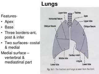
Lungs • The right lung is normally a little larger than the left lung because the middle mediastinum, containing the heart, bulges more to the left than to the right. • Each lung has a half-cone shape, with a base, apex, two surfaces and three borders. • 1. Base • 2. Apex • 3. The two surfaces: Costal surface,mediastinal surface • 4. Three borders: Inferior border , Anterior and Posterior borders
Hilum of the lung • 1.Pulmonary artery • 2. Two pulmonary veins • 3. Main bronchus • 4. Bronchial vessels • 5. Nerves and lymphatics.
Right lung • The right lung has three lobes and two fissures. • Fissures • 1. Oblique fissure • 2. Horizontal fissure
Anatomy of the right lung displaying its two main fissures the horizontal and the oblique
Medial surface of the right lung • 1. Heart • 2.Inferior vena cava • 3.Superior vena cava • 4.Azygos vein • 5.Esophagus
Left lung • The left lung is smaller than the right lung and has two lobes separated by an oblique fissure. • On the anterior surface of the lower part of the superior lobe a tongue-like extension (the lingula of left lung) projects over the heart bulge.
Anatomy of the left lung displaying its fissure that separates it into two lobes upper and lower lobes
Medial surface of the left lung • 1.Heart • 2.Aortic arch • 3.Thoracic aorta • 4. Esophagus
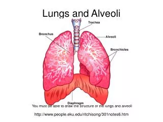
Trachiobronchial Tree • Trachea • Bronchi • Right and left [primary] • Lobar [secondary] [3 or 2] • Segmental [Tertiary] [10] • Bronchiole • Terminal • Respiratory • Alveoli • Alveolar duct • Alveolar Sac • Alveoli
Bifurcation of the trachea • Trachea bifurcates→ two main stem bronchi, right and left • Carina- keel-like ridge between two openings of main stem bronchi • Main stem bronchus divides into lobar bronchi • 3 lobar bronchi on right: upper, middle, and lower • 2 lobar bronchi on left: upper and lower • Each lobar bronchus branches into segmental bronchi that supply a bronchopulmonary segment
The bifurcation of the trachea as seen through an operating bronchoscope
Bronchopulmonary segments • A bronchopulmonary segment is the area of lung supplied by a segmental bronchus and its accompanying pulmonary artery branch.
Bronchopulmonary segment • A bronchopulmonary segment • Is a pyramidally shaped section of lung with its base covered by visceral pleura • Is separated from adjacent segments by connective tissue septa • Is aerated by segmental bronchus • Has its own segmental bronchus and segmental branch of pulmonary artery and segmental branch of bronchial artery but not pulmonary vein • Pulmonary veins are intersegmental
Bronchopulmonary Segment • Has its own Bronchus • Has its own Pulmonary artery (Blue) • Drains to multiple pulmonary veins (Red) between segments • So, each segment has its own bronchus and artery but not its own vein
Aspiration of Foreign Bodies • More likely to enter in right bronchus • Because right bronchus is widerand shorter and runs more vertically than left bronchus • Encountered by dentists • Aspiration of piece of tooth, filling material, or a small instrument
Vasculature of lungs • 2 sets of Blood Supply • 1.Pulmonary Vessels: for Gas Exchange • 2. Bronchial Vessels: for blood supply to lung substance like any other organ
Pulmonary Vessels • Pulmonary artery • Carries unoxygenated blood from heart to lungs • Each artery gives lobar and segmental arteries • Pulmonary veins • Intrasegmental veins drain to intersegmental veins in pulmonary septa, which drain to two pulmonary veins for each lung • Carry oxygenated blood from lungs to heart
Bronchial arteries • Bronchial arteries • Basically supply lung substance • From thoracic aorta • Carry oxygenated blood to tissue of lungs, traveling along posterior surface of bronchi • Left bronchial arteries- arise from thoracic aorta • Right bronchial artery- arise from 3rd posterior intercostal A.
Bronchial veins Drain to azygos and accessory hemiazygos veins
Innervation of lungs • Via pulmonary plexuses • Located anterior and posterior to lung roots • Plexus contains both sympathetic and parasympathetic fibers [2 types of autonomic fibers]
Sympathetic fibers • Innervate smooth muscle of bronchial tree, pulmonary vessels, and glands of bronchial tree • Bronchodilators, vasoconstrictors, and inhibit glandular secretion • Parasympathetic fibers • Preganglionic parasympathetic fibers from vagus nerve • Postganglionic parasympathetic nerves • Innervate smooth muscle of bronchial tree, pulmonary vessels, and glands of bronchial tree • Bronchoconstrictors, vasodilators, and secretomotor to glands
Lymphatic drainage • Lymph from lungs drains to • Pulmonary lymph nodes(along lobar bronchi) • Bronchopulmonary lymph nodes(along main stem bronchi) • Superior and inferior tracheobronchial lymph nodes(superior and inferior to bifurcation of trachea) • Right and left bronchomediastinal trunks • Thoracic duct [left side] and Right lymphatic duct [right side]