Download

1 / 35
350 likes | 489 Vues
Immunologic mechanisms of renal diseases. C hen weilin , PH.D Institute of Immunology, ZJU Email : cwl@zju.edu.cn. Antigens. The cause of immunologically mediated renal disease is antigenic triggering of an immune reaction.

E N D
Immunologic mechanisms of renal diseases Chen weilin,PH.D Institute of Immunology, ZJU Email:cwl@zju.edu.cn
Antigens • The cause of immunologically mediated renal disease is antigenic triggering of an immune reaction. • The list of associated antigens is extensive and continually expanding. These antigens are categorized as renal or non-renal and as self or foreign . • The causative antigen is often unknown.
ANTIGENS ASSOCIATED WITHIMMUNOLOGICALLY MEDIATED RENAL DISEASE
Antigens • To cause immunologically mediated renal disease, an antigen must localize to the kidney and trigger a local immune inflammatory response. • Renal antigens are inherently localized, being constituent proteins of the kidney. • Non-renal antigens require a mechanism for depositing in the kidney.
Immunologic mechanisms of renal diseases • Type II hypersensitivity (Cytotoxic Antibody-mediated ) • Type III hypersensitivity (Immune Complex-mediated ) • Cell-mediated immunity (CD4+,CD8+ T) • Abnormal Immune regulation (Ts) • Immune hereditary factors (HLA)
Type II hypersensitivity Ags on the surface of target cells ↓ body→IgG, IgM ↓ 1. damage the target cell 1) activation of complement 2)opsonization FcR C3bR 3) ADCC 2. target cell dysfunction
Cytotoxic Antibody-mediated Renal Disease • Prototype: Anti-GBM disease (Goodpasture's disease) • Renal damage is caused by linear deposition of antibody specific for type IV collagen of the GBM. The antibody attaches to its antigen and activates the complement. • Cytotoxic antibody localizes along the GBM in a linear pattern with C.
Type III hypersensitivity Ag→body→IgG, IgM, IgA ↓ immune complexes (IC) ↓ soluble IC ↓ ICs are deposited from the circulation into vascular basement membranes ①↓ ② ↓FcR activation of complement plat. and basophils ↓ C3a, C5a →mast cell → release of vasoactive amines ↓ basophils ③ Neutrophils vasodilation ↓ lysosomal edema enzymes→damage the tissue
Immune Complex-mediated Renal Disease • Planted antigen attracts its antibody from the circulation, and a local immune complex is formed. • Immune complex localizes in the mesangium, glomerular capillary wall, or renal interstitium as a lumpy-bumpy pattern. • Small immune complexes are less likely to be deposited, and large immune complexes are preferentially removed by RES minimizing localization in the kidney. • As circulating immune complexes are formed and antibody production increases, the size of the circulating immune complex increases: • removal from the circulation by RES cells or • localization in the mesangium or glomerular capillary wall. • Various endogenous and exogenous substances may function as antigen in immune complex formation. • endogenous nuclear proteins in DNA-anti-DNA IC in lupus nephritis, streptococcal cell wall antigens in post-streptococcal glomerulonephritis.
‘Bumpy’ appearance of immune complexes deposited in the glomerulus in SLE
ACUTE NEPHRITIC SYNDROME(Acute Glomerulonephritis; Postinfectious Glomerulonephritis) • The prototype of an acute nephritic syndrome is poststreptococcal glomerulonephritis (PSGN) due to infection with certain nephritogenic strains of group A -hemolytic streptococci, such as type 12 (associated with pharyngitis) and type 49 (associated with impetigo). • Immunofluorescence microscopy usually shows immune complex deposition with IgG and C in a granular pattern. • The presenting manifestations range from asymptomatic hematuria (in about 50%) and mild proteinuria to full-blown nephritis with gross or microscopic hematuria proteinuria, oliguria, edema, hypertension, and renal insufficiency.
IgA NEPHROPATHY • Berger's disease is now used to describe any idiopathic IgA nephropathy • Patients have gross or microscopic hematuria, often with high blood pressure. The disease usually runs a chronic, slowly progressive course • Mesangial and focal-segmental proliferation and sclerosis may be seen by light microscopy. In bad cases, crescents develop. • Immunofluorescence shows IgA deposited in the mesangium (often with IgG, IgM, and/or C3, but no C4, i.e., the alternate pathway of complement is being activated.) • Serum IgA is often elevated, and IgA-containing immune complexes are often demonstrable, whether or not there is some primary disease to explain their presence
Cell-mediated Renal Disease • The prototype is the renal transplant. • In nearly all non twin transplants, the kidney presents nonself antigens that trigger an immune (predominantly cell-mediated) response. • If the host has been presensitized to antigens of the renal graft, transplantation may trigger hyperacute rejection , resulting in acute renal ischemia, infarction, and transplant loss. • Cell-mediated renal disease appears to play a part in chronic poststreptococcal glomerulonephritis (PSGN). Lymphocytes stimulated by exposure to streptococcal wall antigens may cross-react with renal glomerular antigens, resulting in progressive cell death and sclerosis of the renal parenchyma.
TNF, NO2 IL2, TNF, IFN( IL2, IL4, IL5 lysis IL2, IFN( Mechanisms of graft rejection Inflammation ADCC lysis rejection
Immune hereditary factors • PSGN has been associated with HLA-B12 • IgA nephropathy with HLA-B35 and HLA-DR4 • Anti-GBM or Goodpasture's syndrome with HLA-DR2 • IMN(idiopathic membeanous nephropathy)with HLA- II(DR3、DR2、DQ2、DQ1) • Minmal change nephrosis with HLA-DR7、DR9
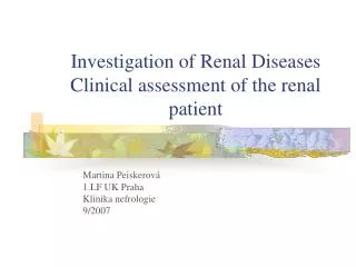
Diagnosis • Renal biopsy and light microscopic examination of stained tissue provide the best method for diagnosing immunologically mediated renal disease, assessing prognosis, and selecting treatment. • Iimmunofluorescence microscopy using fluorescein-labeled specific antibodies often is also helpful in characterizing the type and location of immune components in the kidney. • The type and pattern of C deposition help diagnosis. C deposition usually follows the pattern of immune complex or immunoglobulin deposition or both. However, C3 deposition in the absence of immunoglobulin, Clq, or C4 deposition may occur via alternative pathway activation in type II MPGN..
Urinalysis • Examining the urine for protein and formed elements is often useful. • Nephrotic syndrome is present in virtually all forms of immunologically mediated renal disease. • Abundant protein and frequently lipid-laden modified tubular epithelial cells are found in the urine. • Nephrotic-range proteinuria usually suggests an underlying immune mechanism, although nephrotic syndrome may occur in nonimmune renal disease (eg, diabetes mellitus). • Injury resulting in necrosis, as in acute cytotoxic-type injury of anti-GBM disease, causes significant hematuria. • Immune complex-type injury is associated with hematuria and RBC casts.. • MPGN and membranous glomerulonephritis are associated with significant proteinuria; MPGN usually produces hematuria, but membranous glomerulonephritis rarely does. Minimal-change disease and focal sclerosing glomerulonephritis may produce only proteinuria.
Serologic Analyses • Detect • cytotoxic antibodies in type II renal disease (eg, anti-GBM antibodies, anti-HLA antibodies). • CIC may be found in various immune complex-mediated renal diseases. • Circulating ANCA can be detected in ANCA-mediated renal disease . • Altered levels of C proteins often differentiate types of immunologically mediated renal disease. • When alternative pathway activation predominates (eg, in MPGN and frequently PSGN), C consumption begins with activation of C3; thus, early components of C (Clq, C4, and C2) are not depressed. • In classic pathway activation (eg, in SLE), consumption begins with the early components, which are thereby depressed. • The presence of C3 nephritic factor with depressed C3 but normal Clq, C4, and C2 is virtually diagnostic of MPGN with alternative pathway activation. • Other helpful serologic analyses include: • rising antibody titers to streptococcal antigens in PSGN. • Other postinfective glomerulonephritides eg, a positive test for syphilis, hepatitis-associated antigen, or rising antibody titers to other infective organisms..
Histocompatibility testing • May help diagnose some forms of immunologically mediated renal disease. For example, • PSGN has been associated with HLA-B12, • IgA nephropathy with HLA-B35 and HLA-DR4, and • Anti-GBM or Goodpasture's syndrome with HLA-DR2.