Download
1 / 17
200 likes | 455 Vues
Anatomy. Chapter 8 – Special Senses. Special Senses :. Humans are responsive creatures. “Irritants” are the stimuli that continually enter our nervous system for integration, and may elicit a response. .

E N D
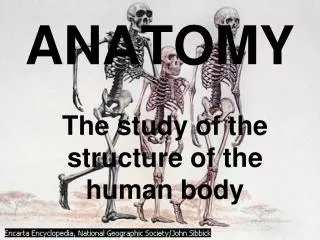
Anatomy Chapter 8 – Special Senses
Special Senses: Humans are responsive creatures. “Irritants” are the stimuli that continually enter our nervous system for integration, and may elicit a response. We are usually told we have five senses that keep us in touch with what is going on in the external world: taste, sight, smell, touch, and hearing . Touch is a mixture of general senses – temperature, pressure, and pain receptors of the skin and proprioceptors of muscle and joints. Smell, sight, taste, and hearing are called special senses. Special sense receptors are either large, complex sensory organs (eyes, ears) or localized clusters of receptors (taste buds and olfactory epithelium).
Frontal bone of orbital The Eye and Vision The adult eye is a sphere about 1 inch (2.5cm) in diameter. The eye is enclosed and protected by a cushion of fat and the bony walls of the orbit. Accessory organs: Eyelids Eyelashes Tarsal glands Conjunctiva Lacrimal apparatus Lacrimal glands Lacrimal canal Lacrimal sac Nasolacrimal duct Lysozyme 70% of sensory input comes through the eyes.
Extrinsic muscles of the Eye Lateral view of right eye Superior view of the right eye Six extrinsic or external, eye muscles attach to the outer surface of the eye. These muscles produce gross movements in the eye allowing the eye to follow objects. The muscles control the reflexive movements of convergence, the movement of the eyes medially when we view close objects. These muscles lose elasticity as we age (40’s).
Internal anatomy of the Eye • The eye is commonly called the eyeball. • The wall is composed of three tunics or coats: • Sclera – outermost tunic • Cornea • Choroid – middle tunic • Pupil • Iris • Retina – inner tunic • Photoreceptors • Optic disc • Fovea centralis Lens focuses light on the retina. It is a flexible, biconvex crystal-like structure. Lens divides the eye into segments or chambers – anterior aqueous humor and posterior vitreous humor.
Pathway of light through the eye and light refraction When light passes from one substance to another , with different densities, its speed changes and its rays are bent, or refracted. Light rays are bent by the cornea, aqueous humor, lens, vitreous humor , before they encounter the retina. The refracting power of the lens can vary by being more or less convex, allowing light to be properly focused on the retina. The eye is set for distance vision – light comes in a parallel waves. Close objects tend to cause light to scatter and diverge (spread out). This causes the lens to bulge to make vision possible, called accommodation. Relative convexity of lens focusing on objects.
Visual Fields and Visual Pathways to the Brain Optic chiasma – the fibers from the medial side of each eye cross over to the opposite side. The fiber tracts that result are the optic tracts. The optic tract fibers synapse with neurons in the thalamus which runs to the occipital lobe of the brain. • Eye Reflexes: • Photopupillary reflex • Accommodation Pupillary reflex • Convergence The visual fields overlap considerably (area of binocular vision).
Errors of refraction and Eye Dysfunction Ophthalmoscope is an instrument used to illuminate the interior of the eye. Nearsighted and Farsighted Fundus of the eye, posterior wall. Night blindness – vitamin A deficiency Color blindness – genetic, x linked Glaucoma – increase pressure in the eye Cataracts appear as a milky structure the seems to fill the pupil.
The Ear: Hearing and Balance Outer Ear: Pinna – Auricle External auditory canal Ceruminus glands - cerumin Tympanic membrane Middle Ear: Ossicles (3) Auditory tube Inner Ear: Cochlea Vestibule Semicircular canals The bony labyrinth of the inner ear is filled with a plasma-like fluid called perilymph. Suspended in the bony labyrinth are membranous sacs that contain a thicker fluid called endolymph.
Mechanisms of Equilibrium: The equilibrium sense responds to the various movements of our head. The equilibrium receptors of the inner ear, the vestibular apparatus, can be divided into two functional groups - static equilibrium and dynamic equilibrium. • Static Equilibrium: • Maculae • Otolithic membrane • Otoliths • Vestibular nerve When the head is tipped, the maculae are stimulated by movement of otoliths in the otolithic membrane with gravitational pull, which creates pull on the hair cells
Dynamic Equilibrium: receptors respond to angular or rotatory movements of the head. The semicircular canals are oriented in the three planes of space, so regardless of which plane one moves in, there will receptors to detect the movement. • Semicircular canals • Cristaampullaris • Cupula • Vestibular nerve Sight and proprioceptors of the muscles and tendons provide information used to control balance to the cerebellum. Ampulla contains a cristaampillaris. Three spatial planes. Head position changes.
Mechanisms of Hearing: Within the cochlear duct is the organ of Corti, which contains the hearing receptors or hair cells. The chambers above and below the cochlear duct contain perilymph. Sound waves reach the cochlea by way of the eardrum, ossicles, and oval window. The transfer of waves sets the cochlear fluids in motion. The receptor cells on the basilar membrane, in the organ of Corti, are stimulated when their “hairs” are bent by the movement of the gel-like tectorial membrane. Once stimulated, the hair cells transmit impulses along the cochlear nerve to the auditory cortex in the temporal lobe. The sound is interpreted and hearing occurs. Sound reaches both ears at different times giving us “stereo”. Cross-sectional view of the cochlea showing the organ of Corti in the cochlear duct. Detailed structure of the organ of Corti.
Hearing and equilibrium deficits: Otitis media – ear infection Fungus in the ear Normal ear drum Perforated ear drum Swimmers ear Tubes in the ear
Deafness – hearing loss of any degree; slight to total inability to hear sound. Conduction deafness - temporary or permanent; something interferes with conduction of sound vibrations in the inner ear – earwax, fusion of ossicles, ruptured eardrum, otitis media. Sensorineural deafness – degeneration or damage to the receptor cells of the organ of Corti, cochlear nerve, neurons or auditory cortex; listening to loud or extended sounds. Meniere’s disease – may be caused by arteriosclerosis, degeneration of cranial nerve VIII, increased pressure of inner ear fluids - progressive deafness. Vertigo – sensation of spinning; nausea. Misshapen stapes
Chemical Senses: Taste and Smell The receptors for taste and smell are classified as chemoreceptors. These receptors respond to chemicals. There are 5 identified taste receptors. The olfactory receptors are sensitive to a much wider range of chemicals. The sense of smell and taste complement each other and will respond to some of the same stimuli. There are 1000’s of olfactory receptors for the sense of smell that occupy a postage stamp-sized area in the roof of each nasal cavity. Our sense of smell is far less acute than other animals. Olfactory receptor cells are neurons equipped with olfactory hairs. The hairs are stimulated by chemicals dissolved in the mucus. The hairs transmit impulses along the olfactory filaments making up the olfactory nerve. Olfactory impressions can be long-lasting and form permanent memories and emotions. Grandma’s house, a vacation.
Taste buds ad the sense of taste: • We have 10,000 taste buds, most are of which are found on the tongue. • Papillae – small peg-like projections on the surface of the tongue • Filiform – sharp • Fungiform - rounded • Circumvallate The cells that respond to the chemicals dissolved in the saliva are epithelial cells called gustatory cells. There are 5 basic tastes: Sweet – Salt – metal ions (found on the tip) Sour – Bitter – alkaloids (posterior); protection – gag reflex Umami – protein, glutamate (beef steak taste)
Influences on taste: taste and smell influence each other • Smell – olfactory receptors • Temperature – • Texture - • Developmental aspects of senses: • Senses form early, the eye by the forth week. The eye functions at birth but is the last to completely develop – learning involved. • Strabismus – congenital eye disorder; “cross-eyed”, can be corrected. • Infants have poor visual acuity – lack complete color vision until 5; depth perception is present. Aging – Presbyopia – lens elasticity is less, difficult to focus on close objects (40 yrs) Atherosclerosis – hardening of arteries in the eye; decrease oxygen and nutrients Decrease in olfactory and gustatory receptors – taste and smell diminishes: 80 years and older; cannot smell and taste, highly season food; lose appetite.