Download

1 / 22
290 likes | 601 Vues
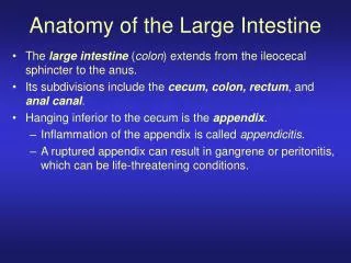
ANATOMY OF THE LARGE INTESTINE. Prof . Ahmed Fathalla Ibrahim Professor of Anatomy College of Medicine King Saud University E-mail: ahmedfathala@hotmail.com. OBJECTIVES. At the end of the lecture, students should: List the different parts of large intestine.

E N D
ANATOMY OF THE LARGE INTESTINE Prof. Ahmed Fathalla Ibrahim Professor of Anatomy College of Medicine King Saud University E-mail: ahmedfathala@hotmail.com
OBJECTIVES At the end of the lecture, students should: • List the different parts of large intestine. • List the characteristic features of colon. • Describe the anatomy of different parts of large intestine regarding: the surface anatomy, peritoneal covering, relations, arterial & nerve supply.
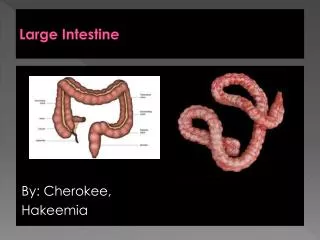
PARTS OF LARGE INTESTINE • CECUM • APPENDIX • ASCENDING COLON • TRANSVERSE COLON • DESCENDING COLON • SIGMOID COLON • RECTUM • ANAL CANAL ABDOMEN PELVIS PERINEUM
CHARACTERISTICS OF COLON(NOT FOUND IN RECTUM & ANAL CANAL) • Teniae coli: 3 longitudinal muscle bands • Sacculations (haustra):teniae coli are shorter than large intestine • Epiploic Appendices : short peritoneal fold filled with fat
PERITONEAL COVERING • PARTS WITH MESENTERY: • Transverse colon • Sigmoid colon • Appendix • Cecum • RETROPERITONEAL PARTS: • Ascending colon • Descending colon • Upper 2/3 of rectum • PARTS DEVOID OF PERITONEAL COVERINGS: • Lower 1/3 of rectum • Anal canal
SURFACE ANATOMY Left hypochondrium Right hypochondrium Epigastrium Right lumbar region Umbilical region Left lumbar region 2/3 McBurney’s point 1/3 ASIS Hypogastrium Left iliac fossa Right iliac fossa
APPENDIXSurface Anatomy The base of appendix is marked by Mc’Burney’s point: A point at the junction of lateral 1/3 & medial 2/3 of a line traced from right anterior superior iliac spine to umbilicus
APPENDIX Opening: at posteromedial aspect of cecum, 1 inch below ileo-cecal junction Positions: 1.Retrocecal: most common 2.Pelvic 3.Subcecal 4.Preilieal 5.Postileal: least common
RELATION BETWEEN ORIGIN & SUPPLY • Origin: Midgut (endoderm) • Artery: Superior Mesenteric • Nerve: Autonomic: • Sympathetic + vagus • Origin: Hindgut (endoderm) • Artery: Inferior Mesenteric • Nerve: Autonomic: • Sympathetic + pelvic splanchnic nerves Left 1/3 Right 2/3 • Origin: ectoderm • Artery: inferior rectal • Nerve: Somatic: inferior rectal Lower part of anal canal
RELATION BETWEEN EMBRYOLOGICAL ORIGIN OF GUT & ITS ARTERIAL SUPPLY
VENOUS DRAINAGE OF GUT • Veins draining gut form the portal circulation • All veins finally end into portal vein which enters the liver
CECUM – ASCENDING & DESCENDING COLONS (ANTERIOR RELATIONS) • Coils of small intestine • Greater omentum • Anterior abdominal wall
1 2 3 5 4 6 1: Iliohypogastric nerve; 2: Ilioinguinal nerve; 3: lateral cutaneous nerve of thigh 4: Femoral nerve; 5: Genitofemoral nerve; 6: Obturator nerve P.M.= psoas major; Q.L.=quadratuslumborum; I.=iliacus; T.A.= transversusabdominis; I.V.C.=inferior vena cava; A.A.=abdominal aorta
CECUM – ASCENDING & DESCENDING COLONS (POSTERIOR RELATIONS) • Cecum: • Psoas major • Iliacus • Ascending colon: • Iliacus • Quadratuslumborum • Descending colon: • Left kidney • Quadratuslumborum • Iliacus • Psoas major
RALATIONS OF TRANSVERSE COLON Anterior: greater omentum, anterior abdominal wall Superior:liver, gall bladder, stomach Inferior: coils of small intestine Posterior:2nd part of duodenum, pancreas
COLIC FLEXURES Position: higher Angle: more acute Hepatic flexure Splenic flexure
RECTUM • Beginning: as a continuation of sigmoid colon at level of S3. • Termination: continues as anal canal, one inch below & in front of tip of coccyx. Its end is dilated to form the rectal ampulla. • Length: 13 cm(5 inches)
RELATIONS OF RECTUM IN PELVIS MALE PELVIS Anterior: seminal vesicles, posterior surfaces of urinary bladder & prostate gland Posterior: sacrum & coccyx FEMALE PELVIS Anterior: posterior wall of vagina Posterior:sacrum & coccyx R R
QUESTION 1 • In which one of the following regions lies McBurney’s point? • Right iliac fossa • Hypogastrium • Right lumbar region • Umbilical region
QUESTION 2 • Which one of the following parts of large intestine is found in the pelvis? • Transverse colon • Anal canal • Rectum • Cecum