Download
1 / 77
810 likes | 859 Vues
Disease of Spine and Spinal Cord. THE SPINAL CORD. The Spinal Cord is an extension of the brain; a thick bundle of nerve fibres. takes a message from the brain, to the muscles to communicate movement messages to the brain, communicating the sense of touch, pain, pressure of heat and cold.

E N D
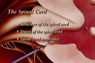
THE SPINAL CORD • The Spinal Cord is an extension of the brain; a thick bundle of nerve fibres. • takes a message from the brain, to the muscles to communicate movement • messages to the brain, communicating the sense of touch, pain, pressure of heat and cold
anterior spinothalamic tract (or ventral) transmits crude touch. lateral corticospinal tract controls movement of contralateral limbs
Lateral corticospinal tract:Movement Lateral spinothalamic tractPain and Temperature
General view • Spinal Cord Injury is damage to the spinal cord that results in a loss of function such as mobility or feeling. Frequent causes of damage are trauma and disease. • Spinal Cord is the major bundle of nerves that carry impulses to/from the brain to the rest of the body. • Spinal Cord is surrounded by rings of bone-vertebra. They function to protect the spinal cord.
Prognosis • Patients with a complete cord injury have a less than 5% chance of recovery. If complete paralysis persists at 72 hours after injury, recovery is essentially zero. • The prognosis is much better for the incomplete cord syndromes. • If some sensory function is preserved, the chance that the patient will eventually be able walk is greater than 50%. • Ultimately, 90% of patients with SCI return to their homes and regain independence. • In the early 1900s, the mortality rate 1 year after injury in patients with complete lesions approached 100%. Much of the improvement since then can be attributed to the introduction of antibiotics to treat pneumonia and urinary tract infection. • Currently, the 5-year survival rate for patients with a traumatic quadriplegia exceeds 90%. The hospital mortality rate for isolated acute SCI is low.
Upper vs. Lower Motor Neuron spasticity, muscle weakness, exaggerated reflexes, and extensor plantar response known as the Babinski sign. • Upper motor neuron lesion • Motor cortex • internal capsule • brainstem • spinal cord • Lower motor neuron lesion • Anterior horn cell • nerve root • plexus • peripheral nerve • neuromuscular junction and muscle decreased tone, decreased strength and decreased reflexes in affected areas
Basic Features of Spinal Cord Disease • UMN findings below the lesion • Hyperreflexia and Babinski’s • Sensory and motor involvement that localizes to a spinal cord level • Bowel and Bladder dysfunction common
Scale of Motor Strength in SCI • The American Spinal Injury Association: • 0 - No contraction or movement • 1 - Minimal movement • 2 - Active movement, but not against gravity • 3 - Active movement against gravity • 4 - Active movement against resistance • 5 - Active movement against full resistance • Assessment of sensory function helps to identify the different pathways for light touch, proprioception, vibration, and pain. Use a pinprick to evaluate pain sensation
Types of Spinal Cord Paralysis • Depending on the location and the extent of the injury different forms of paralysis can occur. • Monoplegia- paralysis of one limb • Diplegia- paralysis of both upper or lower limbs • Paraplegia- paralysis of both lower limbs, Injury in the the thoracic, lumbar, or sacral segments, including the cauda equina and conus medullaris • Hemiplegia- paralysis of upper limb, torso and lower leg on one side of the body • Tetraplegia(Quadraplegia)- paralysis of all four limbs
History • Onset • Acute, subacute, chronic • Symptoms • Pain • Weakness • Sensory • Autonomic • Past history • Family history
Motor Exam • Strength - helps to localize the lesion • Upper cervical • Quadriplegia with impaired respiration • Lower cervical • Proximal arm strength preserved • Hand weakness and leg weakness • Thoracic T1 • Paraplegia • Can also see paraplegia with a midline lesion in the brain • Tone • Increased distal to the lesion
Sensory Exam • Establish a sensory level • Dermatomes • Nipples: T4-5 • Umbilicus: T9-10 • Posterior columns • Vibration • Joint position sense (proprioception) • Spinothalamic tracts • Pain • Temperature
Autonomic disturbances • Neurogenic bladder • Urgency, incontinence, retention • Bowel dysfunction • Constipation more frequent than incontinence • With a high cord lesion, loss of blood pressure control • Alteration in sweating
Investigation of Spinal Cord Disease • Radiographic exams • Plain films • Myelography • CT scan with myelography • MRI • Spinal tap • If you suspect: inflammation, MS, rupture of a vascular malformation
Traumatic Spinal Cord Disease • 10,000 new spinal cord injuries per year • MVA, sports injuries the most common • Victims under 30 yrs old, male>>females • Fx/dislocation of vertabrae most likely to occur at: • C5,6 • T12, L1 • C1,2
Vertebralcolumninjury, withorwithoutneurologicaldeficits, mustalways be soughtandexcluded in a patientwith Multipletrauma. Anyinjuryabovetheclavicle Spineinjury Cervicalspine 55% Thoracicspine 15% Thoracolumbarjunction 15% Lumboscaralspine 15%
Spinal cord injury (SCI) is an insult to the spinal cord resulting in a change, either temporary or permanent, in its normal motor, sensory, or autonomic function.
Excessive manipulation and inadequate immobilization of a patient with a spinal cord injury can cause additional neurological damage and worsen the patient’s outcome.
SPINAL CORD INJURIES Caused by sudden trauma
Incidence: • Race • Whites at 66.4%, AfricanAmericans at 21.1%, Hispanics at 8.8%, Asians at 1.6%, NativeAmericans at 1.1%, andothers at 1%. • Sex • Themale-to-femaleratio of individualswith SCI is 4:1 • Age • 50% ,aged 16-30 years. • Traumatic SCI < 40 years, • Nontraumatic SCI > 40 years.
Associated injuries • Bone fractures (29.3%), • Loss of consciousness (17.8%), • Traumatic brain injury affecting emotional/cognitive functioning (11.5%).
Mechanism: • Destruction from direct trauma • Compression by bone fragments, hematoma, or disk material • Ischemia from damage or impingement on the spinal arteries • Edema could ensue subsequent to any of these types of damage. The different clinical presentations of the above causes of tissue damage are explained further below.
Spinal Shock • An immediate loss of reflex function, called areflexia, below the level of injury • Signs: • Slow heart rate • Low blood pressure • Flaccid paralysis of skeletal muscles • Loss of somatic sensations • Urinary bladder dysfunction • Spinal shock may begin within an hour after injury and last from several minutes to several months, after which reflex activity gradually returns
Neurogenic shock • Neurogenic shock is manifested by the triad of hypotension, bradycardia, and hypothermia. • Shock tends to occur more commonly in injuries above T6, secondary to the disruption of the sympathetic outflow from T1-L2 and to unopposed vagal tone, leading to decrease in vascular resistance with associated vascular dilatation. • Neurogenic shock needs to be differentiated from spinal and hypovolemic shock. Hypovolemic shock tends to be associated with tachycardia
Spinal Shock • An immediate loss of reflex function, called areflexia, below the level of injury • Signs: • Slow heart rate • Low blood pressure • Flaccid paralysis of skeletal muscles • Loss of somatic sensations • Urinary bladder dysfunction • Spinal shock may begin within an hour after injury and last from several minutes to several months, after which reflex activity gradually returns
Classification • The extent of injury is defined by the ASIA Impairment Scale (modified from the Frankel classification), using the following categories: • A - Complete: No sensory or motor function is preserved in sacral segments S4-S5. • B - Incomplete: Sensory, but not motor, function is preserved below the neurologic level and extends through sacral segments S4-S5. • C - Incomplete: Motor function is preserved below the neurologic level, and most key muscles below the neurologic level have muscle grade less than 3. • D - Incomplete: Motor function is preserved below the neurologic level, and most key muscles below the neurologic level have muscle grade greater than or equal to 3. • E - Normal: Sensory and motor functions are normal.
Spinal Cord Syndromes can be classified into either complete or incomplete categories • Complete – characterized as complete loss of motor and sensory function below the level of the traumatic lesion • Incomplete – characterized by variable neurological findings with partial loss of sensory and/or motor function below the lesion
Injuries by ASIA classification • Incomplete tetraplegia - 29.5% • Complete paraplegia - 27.9% • Incomplete paraplegia - 21.3% • Complete tetraplegia - 18.5% • The most common neurologic level of injury is C5. In paraplegia, T12 is the most common level.
Functional outcome measures • Several functional-outcome measures are reliable and valid for use in SCI. A common scale for the measurement of functional ability is the Functional Independence Measure (FIM), which uses a 7-point scale to measure 18 items in the following 6 categories: • Mobility • Locomotion • Self-care • Continence of the bowel and/or bladder • Communication • Social cognition • On the FIM scale, a score of 1 indicates total dependence on a caregiver, and a score of 7 indicates independence. Numbers between 1 and 7 represent different levels of assistance required from a caregiver or assistive device to perform a specific skill
Clinical syndromes • Central cordsyndromeoften is associatedwith a cervicalregioninjuryleadingtogreaterweakness in theupperlimbsthan in thelowerlimbswithsacralsensorysparing. • Brown-Séquardsyndromeoften is associatedwith a hemisectionlesion of thecord, causing a relativelygreateripsilateralproprioceptiveand motor losswithcontralateralloss of sensitivitytopainandtemperature. • Anteriorcordsyndromeoften is associatedwith a lesioncausingvariableloss of motor functionandsensitivitytopainandtemperature, whileproprioception is preserved. • Conusmedullarissyndrome is associatedwithinjurytothesacralcordandlumbarnerverootsleadingtoareflexicbladder, bowel, andlowerlimbs, whilethesacralsegmentsoccasionallymayshowpreservedreflexes (eg, bulbocavernosusandmicturitionreflexes). • Caudaequinasyndrome is duetoinjurytothelumbosacralnerveroots in thespinalcanalleadingtoareflexicbladder, bowel, andlowerlimbs.
Central Cord Syndrome • Usually involves a cervical lesion • May result from cervical hyperextension causing ischemic injury to the central part of the cord • Motor weakness is more present in the upper limbs then the lower limbs • More commonly seen in older patients with cervical arthritis or narrowing of the spinal cord
Results from an injury to only half of the spinal cord and is most noticed in the cervical region Motor loss is evident on the same side as the injury to the spinal cord Sensory loss is evident on the opposite side of the injury location (pain and temperature loss) Bowel and bladder functions are usually normal Brown-Sequard Syndrome
Anterior Spinal Cord Syndrome • Compression of the artery that runs along the front of the spinal cord • Patients have a variable amount of motor function below the level of injury • Sensation to pain and temperature are lost while sensitivity to vibration and proprioception are preserved
Conus Medullaris vs. Cauda Equina Lesion FindingConusCE Motor SymmetricAsymmetric Sensory loss Saddle Saddle Pain Uncommon Common Reflexes Increased Decreased Bowel/bladder Common Uncommon
Scale of Motor Strength in SCI • The American Spinal Injury Association: • 0 - No contraction or movement • 1 - Minimal movement • 2 - Active movement, but not against gravity • 3 - Active movement against gravity • 4 - Active movement against resistance • 5 - Active movement against full resistance • Assessment of sensory function helps to identify the different pathways for light touch, proprioception, vibration, and pain. Use a pinprick to evaluate pain sensation.
Motor level - Determined by the most caudal key muscles that have muscle strength of 3 or above while the segment above is normal (= 5) • Motor index scoring - Using the 0-5 scoring of each key muscle with total points being 25/extremity and a total possible score of 100 • Sensory level - Most caudal dermatome with a normal score of 2/2 for both pinprick and light touch • Sensory index scoring - Total score from adding each dermatomal score with possible total score (= 112 each for pinprick and light touch) • Neurologic level of injury - Most caudal level at which both motor and sensory levels are intact, with motor level as defined above and sensory level defined by a sensory score of 2
Zone of partial preservation - This index is used only when the injury is complete. All segments below the neurologic level of injury with preservation of motor or sensory findings • Skeletal level of injury - Level of greatest vertebral damage on radiograph • Lower extremities motor score (LEMS) - Uses the ASIA key muscles in both lower extremities with a total possible score of 50 (ie, maximum score of 5 for each key muscle L2, L3, L4, L5, and S1 per extremity). A LEMS score of 20 or less indicates patients are likely to be limited ambulators. A LEMS of 30 or more suggests that patients are likely to be community ambulators.
Spinal Cord Paralysis Levels C1-C3 • All daily functions must be totally assisted • Breathing is dependant on a ventilator • Motorised wheelchair controlled by sip and puff or chin movements is required C4 • Same as C1-C3 except breathing can be done without a ventilator
C5 • Good head, neck, shoulder movements, as well as elbow flexion • Electric wheelchair, or manual for short distances C6 • Wrist extension movements are good • Assistance needed for dressing, and transitions from bed to chair and car may also need assistance C7-C8 • All hand movements • Ability to dress, eat, drive, do transfers, and do upper body washes
T1-T4 (paraplegia) • Normal communication skills • Help may only be needed for heavy household work or loading wheelchair into car T5-T9 • Manual wheelchair for everyday living • Independent for personal care T10-L1 • Partial paralysis of lower body
L2-S5 • Some knee, hip and foot movements with possible slow difficult walking with assistance or aids • Only heavy home maintenance and hard cleaning will need assistance