Download
1 / 73
730 likes | 1.1k Vues
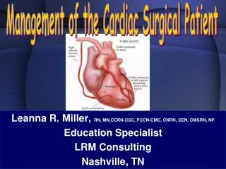
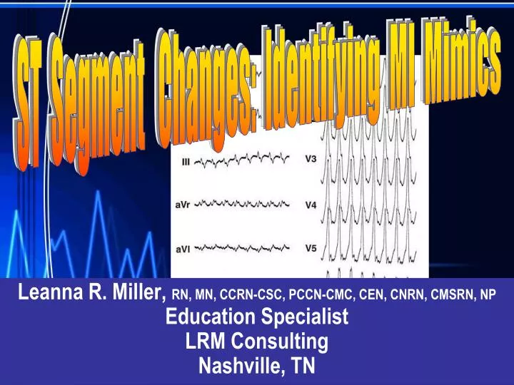
ST Segment Changes: Identifying MI Mimics. Leanna R. Miller, RN, MN, CCRN-CSC, PCCN-CMC, CEN, CNRN, CMSRN, NP Education Specialist LRM Consulting Nashville, TN. ST Segment Changes: Identifying MI Mimics. Objectives Evaluate common abnormalities that mimic myocardial infarction .

E N D
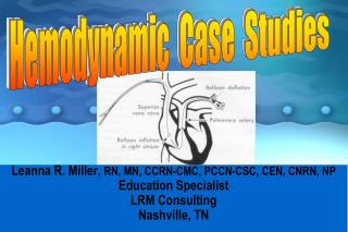
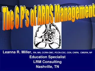
ST Segment Changes: Identifying MI Mimics Leanna R. Miller, RN, MN, CCRN-CSC, PCCN-CMC, CEN, CNRN, CMSRN, NP Education Specialist LRM Consulting Nashville, TN
ST Segment Changes: Identifying MI Mimics • Objectives • Evaluate common abnormalities that mimic myocardial infarction. • Identify the criteria for pericarditis and evidence – based interventions. • Differentiate between pulmonary embolus and myocardial infarction using diagnostic criteria.
ST Segment Changes: Identifying MI Mimics • Acute Coronary Syndromes • Unstable Angina • Non ST segment Elevation MI (NSTEMI) • ST segment Elevation MI (STEMI)
ST Segment Changes: Identifying MI Mimics • Acute Coronary Syndromes • Clinical Symptoms • typical • atypical
ST Segment Changes: Identifying MI Mimics • Acute Coronary Syndromes • Diagnostics • Echocardiography • Lab • ABGs • H & H • enzymes
ST Segment Changes: Identifying MI Mimics • Acute Coronary Syndromes • Diagnostics • ECG (12 or 15 lead) • T wave inversion • ST segment elevation • Q wave • reciprocal ST segment • depression
ST Segment Changes: Identifying MI Mimics Variation to ST – Segment Elevation
ST Segment Changes: Identifying MI Mimics High acute risk factors for progression to myocardial infarction or death • recurrent chest pain at rest • dynamic ST-segment changes: ST-segment depression > 0.1 mV or transient (<30 min) ST-segment elevation >0.1 mV • elevated Troponin-I, Troponin-T, or CK-MB levels
ST Segment Changes: Identifying MI Mimics High acute risk factors for progression to myocardial infarction or death • hemodynamic instability within the observation period • major arrhythmias (ventricular tachycardia, ventricular fibrillation) • early post-infarction unstable angina • diabetes mellitus
ST Segment Changes: Identifying MI Mimics • Acute Pericarditis • Introduction • causes physical discomfort • predisposition to tachydysrhythmias
ST Segment Changes: Identifying MI Mimics • Acute Pericarditis • ECG Criteria • ST segment elevation • PR segment depression • T wave flattening or inversion • atrial dysrhythmias
ST Segment Changes: Identifying MI Mimics • Acute Pericarditis • ST segment elevation • not isolated or discrete segments • upward concavity • may be notching at the junction of • QRS and ST segment • no reciprocal ST segment depression
ST Segment Changes: Identifying MI Mimics • Acute Pericarditis • PR interval • interval between end of P wave and • beginning of QRS may be depressed • most often seen in lead II and V • leads may be only ECG finding
ST Segment Changes: Identifying MI Mimics • Acute Pericarditis • T wave flattening or inversion • no T wave inversion during acute phase • uncomplicated pericarditis: negative • T waves only occur in leads which usually • have negative T waves (aVR & V1)
ST Segment Changes: Identifying MI Mimics • Acute Pericarditis • Atrial dysrhythmias • SVT in postoperative open heart patient • treat with low dose steroids
ST Segment Changes: Identifying MI Mimics • Acute Pericarditis • Complications (pericardial effusion) • dampening of electrical output • low voltage in all leads • ST segment & T wave changes
ST Segment Changes: Identifying MI Mimics • Acute Pericarditis • Complications (pericardial effusion) • freely rotating heart produces • electrical alternans
ST Segment Changes: Identifying MI Mimics • Dressler’s Syndrome • Introduction • postmyocardial infarction syndrome • autoimmune process
ST Segment Changes: Identifying MI Mimics • Dressler’s Syndrome • Clinical Presentation • low – grade fever • chest pain (worsens with deep • breath; lessens with sitting up • and leaning forward) • pericardial friction rub
ST Segment Changes: Identifying MI Mimics • Dressler’s Syndrome • 12 – lead ECG • diffuse ST segment elevation across the precordial leads
ST Segment Changes: Identifying MI Mimics • Dressler’s Syndrome • Treatment • corticosteroid administration • monitor for complications (effusion)
ST Segment Changes: Identifying MI Mimics • Pulmonary Embolus • Introduction • sudden massive PE produces ECG changes • must get 12 – lead to rule out MI
ST Segment Changes: Identifying MI Mimics • Pulmonary Embolus • ECG Findings • RVH with strain • RBBB pattern in V1 • large S wave in Lead I; large Q wave in Lead • III (S1Q3 pattern)
ST Segment Changes: Identifying MI Mimics • Ventricular Aneurysm • Introduction (etiology) • myocardial infarction • congenital • cardiomyopathy • inflammatory • idiopathic
ST Segment Changes: Identifying MI Mimics • Ventricular Aneurysm • Introduction • infereolateral wall of LV • symptoms include CHF & exercise – • induced syncope (VT)
ST Segment Changes: Identifying MI Mimics • Ventricular Aneurysm • ECG Findings • persistent ST segment elevation • small q wave in II, III, & aVF • sustained VT with RBBB morphology
ST Segment Changes: Identifying MI Mimics • Ventricular Aneurysm • Treatment • surgical resection • antidysrhythmics • anticoagulants • treat heart failure • ablation therapy
ST Segment Changes: Identifying MI Mimics • Left Bundle Branch Block (LBBB) • QRS duration > 0.12 second • absence of septal q waves and S wave • in I, aVL, & V5 – 6 (+ complex usually • notched) • broad QS or rS in V1 – 3(- complex)
ST Segment Changes: Identifying MI Mimics • Left Bundle Branch Block (LBBB) • S – T, T wave changes in leads I, • aVL & V5 – 6 (T wave opposite QRS) • delayed intrinsicoid deflection over • left ventricle (V6); normal over V1
ST Segment Changes: Identifying MI Mimics • Left Bundle Branch Block (LBBB) • hypertensive heart disease • aortic stenosis • degenerative changes of the conduction • system • coronary artery disease