Download
1 / 66
660 likes | 803 Vues
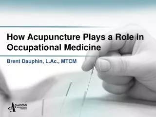
The Role of Physiatry in Occupational Medicine January 31, 2013. James Petros, M.D., Q.M.E. Board-Certified, Physiatry Board-Certified, Pain Medicine Board-Certified, Internal Medicine. A little bit about Dr. Petros…. Curriculum Vitae. What is a Physiatrist?.

E N D
The Role of Physiatry in Occupational MedicineJanuary 31, 2013 James Petros, M.D., Q.M.E. Board-Certified, Physiatry Board-Certified, Pain Medicine Board-Certified, Internal Medicine
A little bit about Dr. Petros… Curriculum Vitae
What is a Physiatrist? • Specialist in physical medicine and rehabilitation (PM&R) • physikos –“physical” iatreia –“art of healing” • Integrates functional medicine, orthopedics, neuroscience, pain management, therapeutic rehabilitation
Nonsurgical Address muscles, nerves, bones, joints, tendons, ligaments Functional restoration “Quality of life”
Origins of Physiatry • Formed in the 1930s to address neuro-musculo-skeletal problems • Grew drastically during WWII • Approved as a specialty of medicine in 1947
Physiatry: The “Broad” Specialty • Pain management • Musculoskeletal • Electrodiagnosis • Spinal cord injury • Traumatic brain injury • Amputation, Orthotics, Prosthetics • General rehab • Sports • Industrial
What Sets Dr. Petros Apart in Work Comp? • Triple-board certification • Diverse skill set • Refined knowledge and experience • UR work • AME/QMEs • Case load • Attention to detail • RTW approach • “Make each visit count” • “A-Z” mindset • Manage and salvage most complicated claims • Resources at Alliance
Other Important factors… • Compassion • Advocacy • Rapport • I love what I do
Conditions Treated • Low back pain • Mid back pain • Neck pain • Herniated disc • Spinal stenosis • Lumbar radiculopathy (sciatica) • Facet syndrome • Sacroiliac dysfunction • Whiplash syndrome • Repetitive stress injuries • Shoulder/elbow/hand problems • Hip/knee/foot problems • Myofascial/muscle pain • Carpal tunnel syndrome • Cubital tunnel syndrome • Neuropathic (nerve) pain • Arthritis • Post-surgical pain • Headaches
My Specialized Skills • Diagnosis • Medication optimization • Therapy • Interpretation of X-rays & MRIs • Peripheral joint, muscle, bursa, tunnel, and nerve injections • Spinal procedures under fluoroscopy • Electrodiagnosis
Spinal Procedures • Epidural Steroid Injections (ESIs) • Facet Medial Branch Blocks • Facet Intra-articular Injections • Facet Radiofrequency Rhizotomy • Sacroiliac Joint Injections • Sympathetic Ganglion Blocks • Piriformis Muscle Injections • Discograms • Trigger point injections
Showcase Epidural Steroid Injections (ESIs) Facet Joint Procedures EMG/NCS
Medical Criteria for ESIs • Persistent radiculopathy • Inflammation related to: • Spinal disc herniation (“discogenic pain”) • Degenerative disc disease • Spinal stenosis
What is Radiculopathy? Spinal condition Compressed nerve root Pain, numbness, tingling, or weakness along the course of the nerve
Risk Factors for Radiculopathy? Excessive or repetitive load on the spine Aging Obesity Smoking Family history
Causes of Radiculopathy? Disk herniation Inflammation from trauma or degeneration Bone spur (osteophytes) Tumor Infection Scoliosis Diabetes
Goals Following ESIs • Reduce pain/tingling/numbness/weakness • Restore range of motion • Facilitate progress in more active treatment programs • Avoid surgery
“Based on the evaluation of multiple randomized and non-randomized trials, transforaminal epidural injections provide strong evidence for short-term and long-term relief.” Manchikanti L, et al. Evidence-based practice guidelines for interventional techniques in the management of chronic spinal pain. Pain Phys 2003;6:3-81. [1175 references].
Showcase Epidural Steroid Injections (ESIs) Facet Joint Procedures EMG/NCS
Facet Mediated Pain “Facet syndrome” 30-50% of cases we see Causes: abnormal spinal loading, trauma, inflammation, degeneration, fracture Diagnosis requires clinical suspicion Focal tenderness over joint Pain exacerbated by extension of spine (closing of facet joints) MRI may be normal Treatment mainstays: education, medications, therapy
Facet Procedures • Diagnostic • Medial branch blocks • Therapeutic • Intra-articular steroid injection • Radiofrequency Ablation (Rhizotomy)
Lumbar Medial Branch Block “Combined evidence of the medial branch blocks from one randomized trial, complimented with two non-randomized trials (one prospective and one retrospective evaluation) provided strong evidence of short-term relief.”: Manchikanti L, et al. Evidence-based practice guidelines for interventional techniques in the management of chronic spinal pain. Pain Phys 2003;6:3-81. [1175 references]”
Showcase Epidural Steroid Injections (ESIs) Facet Joint Procedures EMG/NCS
Electrodiagnosis • Electromyography (EMG) • Nerve conduction studies (NCS)
Originated in 19th century More consistently used over last 30-40 years Extension of physical exam Evaluate integrity of peripheral nervous system
EMG/NCS – FREQUENTLY ASKED QUESTIONS What does the EMG/NCS test? The EMG/NCS study examines the integrity of the peripheral nerves and muscles of the body. The study does NOT examine the brain or spinal cord. It is important to realize that you can have a nerve or muscle problem, even though you may not “think” you have any nerve or muscle problems. This test does NOT measure pain. You may have a normal EMG-NCS study, even though you have severe pain. What are the different parts of the study? The study is usually done in two parts: (1) NCS, and (2) EMG (i.e. “needle” exam). How long is the study? Each EMG/NCS study varies from patient to patient, depending on what results are obtained. As such, the study may take as little as 20 minutes, or as much as 2 hours. What is the Nerve Conduction Study or NCS? The NCS involves examining the nerves in your arms or legs. This consists of attaching wires to the surface of your skin, and administering a small “shock” to see how well the nerves react and function. These results are monitored on a computer. What is the Electromyography or EMG? The EMG examines the muscle activity in your body. This study consists of inserting a sterile, individually wrapped, needle into your various muscles and monitoring their activity. These results are monitored on a computer. You will probably be stuck 5-7 times per arm or leg. There is NO shocking during the EMG. Is the EMG or NCS painful? The “shocks” during the NCS are not painful, although they may produce a tingling sensation. The needle “sticks” during the EMG feels like a small ant bite, and can sometimes be uncomfortable, but not painful.
Utility of EMG/NCS • Establish correct diagnosis • Screen for other conditions • Determine acuity vs. chronicity • Localize lesion • Determine treatment • Prognosticate
When to Consider EMG/NCS? • Pain • Numbness • Tingling • Weakness • Atrophy • Fatigue
Electrodiagnostic Protocol • NCS • Upper: median, ulnar, radial • Lower: tibial, peroneal, sural, superficial peroneal • EMG • Upper: cervical paraspinals, deltoids, biceps, triceps, pronator teres, 1st dorsal interosseous, abductor pollicis brevis • Lower: lumbar paraspinals, gluteus medius, biceps femoris, vastus medialis, tibialis anterior, gastrocnemius
Test Segments NCS EMG (needle) Spontaneous electrical activity Insertional activity Waveform shape Recruitment patterns • Latency • Amplitude • Conduction velocity • Signal quality
Alcoholic neuropathy Amyotrophic lateral sclerosis Axillary nerve dysfunction Becker's muscular dystrophy Brachial plexopathy Carpal tunnel syndrome Centronuclear myopathy Cervical spondylosis Charcot-Marie-Tooth disease Chronic Immune Demyelinating Poly[radiculo]neuropathy (CIDP) Dermatomyositis Duchenne muscular dystrophy Facioscapulohumeral muscular dystrophy Familial periodic paralysis Femoral nerve dysfunction Friedreich's ataxia Guillain-Barre Lambert-Eaton Mononeuropathy Motor neuron disease Multiple system atrophy Myasthenia gravis Myopathy (muscle degeneration, which may be caused by a number of disorders, including muscular dystrophy) Myotubular myopathy Neuromyotonia Peripheral neuropathy Poliomyelitis Polymyositis Radial nerve dysfunction Radiculopathy Sciatic nerve dysfunction Sleep bruxism Spinal stenosis Tibial nerve dysfunction Ulnar nerve dysfunction Examples of Electrodiagnoses
Case Study • 44 y.o. male • Generally healthy • Limousine driver • MVA on the job • Vehicle totaled
Immediate Symptoms • Dazed and confused (no LOC) • Headaches • Neck Stiffness • Generalized soreness
EMS activated on the scene • ER • Non-focal neuro exam • C-spine X-rays (negative) • Head CT scan (negative) • Discharge to home with neck brace, NSAIDs, muscle relaxers, and Vicodin
Next Day • Neck pain (main complaint) • Headaches • Mid back pain • Low back pain • Right knee pain • Patient referred to AOM
AOM Care (Day #3) • Additional x-rays • Thoracic spine: Negative • Lumbar spine: Negative • Right knee: Negative • Diagnoses: • C/T/L strains due to whiplash • Right knee sprain from impact with dashboard
AOM Care (Day #3) • Plan • Physical therapy • HEP • NSAIDs, muscle relaxers • RTC 2 weeks
AOM Care (Day #17) • Mid back pain resolved • Low back pain resolved • Right knee pain resolved • Neck pain worse • RUE paresthesias • Neck ROM decreased • Weak triceps • Right Spurling’s positive
AOM Care (Day #17) • Plan • Add Vicodin • Start chiropractic • No driving • RTC 2-3 weeks
AOM Care (Day #33) Worsening neck pain and headaches Neck pain radiating stronger into RUE Neck ROM still limited Weak elbow extension Right hand dorsal numbness Spurling’s still positive
AOM Care (Day #32) • Plan • Continue medications • Hold therapy • Refer for C-spine MRI • Refer to Physiatry