Download
1 / 25
310 likes | 717 Vues
FETAL CHEST FETAL HEART. FETAL CHEST. DIAPHRAGM Assess diaphragm (thin echogenic line) Diaphragm hernias Lung and bowel similar echogenicity- Look for peristalsis Left easier to see than right due to gastric bubble LUNGS Look for pulmonary masses CCAM Sequestration Pulmonary hypoplasia

E N D
FETAL CHEST DIAPHRAGM Assess diaphragm (thin echogenic line) Diaphragm hernias Lung and bowel similar echogenicity- Look for peristalsis Left easier to see than right due to gastric bubble LUNGS Look for pulmonary masses CCAM Sequestration Pulmonary hypoplasia PLEURA - effusions MEDIASTINUM - masses
CONGENITAL DIAPHRAGM HERNIA Bochdalek - 90% on left; most unilat All should have amniocentesis and dedicated echo Secondary pulmonary hypoplasia is major cause of mortality Findings Polyhydramnios Stomach/bowel/liver adjacent to heart Peristalsis in chest Mediastinal shift Absent gastric bubble Reduced abdominal circumference compared to rest of fetal biometry
Associated anomalies Aneuploidy (T18, T21); NTD; CHD; malrotation, omphalocele DDX CCAM Other cystic masses such as foregut duplication cysts are rare
CCAM Most common fetal lung mass Types I-III Types I and II macroscopic cysts >5mm with good prognosis and hydrops is rare Small risk of malignant degeneration (rhabdomyosarcoma) Imaging Macroscopic types appear cystic Microscopic types appear solid (echogenic) Pulmonary hypoplasia of normal lung - degree determines prognosis Mediastinal shift - cardiac compromise; polyhydramnios (impaired swallowing) Associations (type II) Cardiac anomalies Pulmonary sequestration Pectus excavatum Jejunal atresia Renal agenesis, prune-belly syndrome Pathology Hamartomatous proliferation of terminal bronchioles Cysts lined by respiratory epithelium and communicate with airways at birth
EXTRALOBAR SEQUESTRATION More common in males (4:1) 90% LLL or below diaphragm Always airless as it has its own pleural envelope and no communication with bronchial tree Systemic arterial supply - Aorta 80% Systemic venous drainage - IVC, azygos, portal v Imaging Findings Solid hyperechogenic mass Look for systemic arterial supply on Doppler Polyhydramnios Hydrops Associations 65% CDH Cardiac GI, Renal, Vertebral anomalies Often regress in utero DDX CCAM Congential lobar emphysema (initially filled with fetal fluid) Neuroblastoma
PULMONARY HYPOPLASIA Agenesis – complete absence of one or both lungs (airways, alveoli, and vessels) Aplasia – absence of lung except for a rudimentary bronchus that ends in a blind pouch Hypoplasia – decrease in number and size of airways and alveoli Primary Secondary Bilateral - Oligohydramnios (Potter’s sequence); Skeletal dysplasia Unilateral - CCAM; Sequestration; CDH; Hydrothorax Imaging Reduced thoracic circumference (<2SD) is suggestive Fetal lung maturity best sssessed with lecithin:sphingomyelin ratio in amniotic fluid Echogenic pattern unreliable marker for maturity
PLEURAL EFFUSION = abnormal Fetal hydrops Chromosomal Underlying mass Infection Lymphangiectasia Chylothorax - assoc with T21 and Turner’s MEDIASTINAL MASSES Anterior Medistinum Teratoma Cystic hygroma Normal Thymus Posterior Mediastinum Neurogenic tumours Enteric cyst
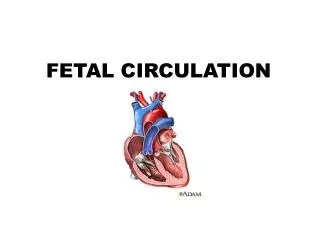
FETAL HEART Technique Abdominal situs view 4-chamber view LVOT Posterior/central to RVOT Runs left to right RVOT Anterior to LVOT Runs right to left Bifurcates early: DA and RPA Check for antegrade flow in DA Anatomical trifurcation: DA, RPA, LPA 3-vessel view amniocentesis indicated in all abnormal: 15-40% will have chromosomal anomalies ventricles/atria are of roughly same size as other ventricle/atria 3 in 1 rule: heart fills 1/3 of axial chest Cardiac circumference 1/2 chest circumference Length atrial septum: ventricular septum 1:2 Normal HR: 120-160bpm, SR
Best seen on Four-Chamber View Septal defect Endocardial cushion defect (AVSD) Hypoplastic left heart Ebstein’s anomaly Critical AS Coarctation
Best Seen on Outflow Tract Views Tetralogy of Fallot Transposition Truncus Arteriosus Pentalogy of Cantrell
Maternal Risk Factors for CHD Diabetes Infection - rubella, CMV SLE Drugs - EtOH, Phenytoin, lithium FHX of heart disease, previous child with CHD Arrhythmia
VSD Most common CHD (1:1000) Membranous 80% vs Muscular 10% vs Outlet (ECD) 5% Don’t mistake membranous to muscular transition for VSD
Endocardial Cushion Defect 40% have Trisomy 21 EC forms lower atrial septum, superior ventricular septum, anterior MV leaflet and septal TV leaflet
Transposition of Great Vessels Aorta arises from RV and pulmonary trunk from LV Aorta and pulmonary artery are parallel instead of perpendicular to each other
Tetralogy of Fallot Tetralogy Infundibular RV outflow tract stenosis Overriding aorta VSD Hypoplastic RV LV and RV are symmetric due to equal pressures Often missed on 4-chamber view
Ebstein’s Anomaly Septal and posterior leaflets of tricuspid valve prolapse and are integrated into RV wall Atrialisation of RV Large RA due to massive regurg Maternal lithium is a risk factor
Pulmonary Atresia Hypoplastic RA and RV Pulmonary artery calibre may be normal Reversed flow in DA
Pericardial Effusion >2mm Associated with hydrops fetalis, congenital infection and cardiac anomalies Look for fluid in other compartments (hydrops) Look for signs of congential infections Cerebral calcification Hepatic calcifciation Echogenic bowel
Endocardial Fibroelastosis Increased echogenicity of endocardium Ventricular dilatation and poor contractility Ectopia Cordis
Rhabdomyoma Hamartoma of myocytes Strong association with Tuberous Sclerosis 50-85% of fetuses with it have TS 50% of TS have it Echogenic mass, usually intraventricular, can arise from IV septum
FETAL ARRHYTHMIAS PAC and PVC common and benign SVT is the most common tachyarrhythmia - CMX hydrops Fetal bradycardia (HR <100 for >10sec) If persistent - consider structural cardiac defects or maternal CVD Fetal heart block 40-50% have structural abnormality - usually lethal Associated with maternal SLE, RA, Scleroderma