Download
1 / 27
320 likes | 619 Vues
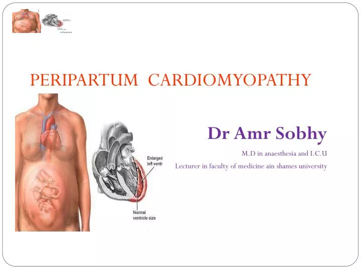
PERIPARTUM CARDIOMYOPATHY Dr Amr Sobhy M.D in anaesthesia and I.C.U Lecturer in faculty of medicine ain shames university. Objectives. Definition Epidemiology Diagnosis Management Update Prognosis. Definition.

E N D
PERIPARTUM CARDIOMYOPATHY Dr Amr Sobhy M.D in anaesthesia and I.C.U Lecturer in faculty of medicine ain shames university
Objectives • Definition • Epidemiology • Diagnosis • Management • Update • Prognosis
Definition • An idiopathic cardiomyopathy presenting with heart failure secondary to LV systolic dysfunction towards the end of pregnancy or in the months following delivery, where no other cause of heart failure is found. It is a diagnosis of exclusion. The LV may not be dilated but the ejection fraction is nearly always reduced below 45% Heart Failure Association of the ESC Working Group on PPCM (Sliwa et al., 2010)
Epidemiology Incidence • The incidence in the west ranges from 1 in 4000 deliveries • 60% present within the first 2 months postpartum • Up to 7% may present in the last trimester of pregnancy. • Geographic variations exist with a higher incidence reported in areas of Africa .
Epidemiology Etiology Still unknown. • Nutritional deficiencies • Small vessel coronary artery abnormality • Hormonal effects • Toxemia • Maternal immunologic response to fetal antigen or myocarditis
Epidemiology Predisposing factors • Maternal age greater than 30 yr • Multiparous • Twinning • Racial origin (black) • Hypertension and eclamptic patients • Nutritional deficiencies • No family history
Diagnosis Criteria 1.Development of Cardiac failure in the last month of pregnancy or within 5 month after delivery 2. Absence of an identifiable cause for the cardiac failure. 3.Absence of recognizable heart disease prior to the last month of pregnancy. 4.Left ventricular systolic dysfunction demonstrated by classic Echo Cardio Graphic criteria such as depressed shortening fraction or ejection fraction. The National Heart, Lung and Blood Institute and the Office of rare diseases (1997)
Diagnosis Criteria 1.Development of Cardiac failure in the last month of pregnancy or within 5 month after delivery 2. Absence of an identifiable cause for the cardiac failure. 3.Absence of recognizable heart disease prior to the last month of pregnancy. 4.Left ventricular systolic dysfunction demonstrated by classic Echo Cardio Graphic criteria such as depressed shortening fraction or ejection fraction. The National Heart, Lung and Blood Institute and the Office of rare diseases (1997)
Diagnosis Clinical Presentation • Symptoms: • Paroxysmal Nocturnal Dyspnea • Dyspnea on Exertion • Cough • Orthopnea • Chest Pain • Abdominal Discomfort • Palpitation • Signs: • Cardiomegaly • Gallop Rhythm • Edema • Holosystolic murmur • Thromboembolic Manifestation
Diagnosis Clinical Presentation • Often unrecognized, as symptoms of normal pregnancy commonly mimic those of mild heart failure. • In the absence of any cardiac symptoms, one of the early indications about this condition is Fetal growth retardation during evaluation of the fetus with a fetal monitor and ultrasound
Diagnosis Investigation 1.Chest X rays • Cardiomegaly with pulmonary oedema • Pulmonary venous congestion. 2. The E.C.G • Nonspecific ST and T wave changes • Atrialor ventricular arrhythmias and • conduction defects
Diagnosis Investigation 3.ECHO • Enlargement of all four chambers with marked reduction in left ventricular systolic function • Small to moderate pericardial effusion • Mitral, tricuspid and pulmonary regurgitation • Ventricular wall motion, ejection fraction and cardiac output are decreased . • Pulmonary wedge pressure is increased.
Management Consultations • Cardiologist • High-risk obstetrician • Anesthesiologist - Neuraxial anesthesia is preferred to avoid myocardial depression from inhaled anesthetics; for this reason, as the mother nears delivery, low-molecular-weight heparin should be used with caution.
Management Vigorous treatment of AHF • Non-pharmacological • Salt restriction (4gm/d) • Water restriction (2 L/D) • Pharmacological • Pre-load reduction (diuretics, nitrates) • After-load reduction (hydralazine, nitrates, amlodipine) • ACE-I contraindicated during pregnancy • Ionotropes (digoxin, dopamine, dobutamine) • Beta-blockers • Anticoagulant
Update in Management Immunosuppressive agents • May be initiated in patients with PPCM and biopsy-proven myocarditis, but efficacy is unclear • Empiric immunosuppression, in the absence of evidence of myocarditis, is not currently recommended • Cardiac MRI could guide the immunosuppressive therapy
Update in Management pentoxifylline
Update in Management Levosimendan
Update in Management Bromocriptine
Update in Management • Since the disease may be reversible, the temporary use of Intra Aortic Balloon Pump or LV assist device may help to stabilize the patient’s condition pending improvement.
Update in Management Cardiac Resynchronization Therapy and Implantable Cardioverter/Defibrillators • LV ejection fraction < 35% persists after 6 months following presentation. • Patients with recurrent symptomatic ventricular arrhythmias • If NYHA III and IV heart failure symptoms and a QRS duration > 120 ms
Update in Management Cardiac Transplantation
Prognosis • Prognosis seems dependent on recovery of left ventricular function. • 30% of patients return to baseline ventricular function within 6 months. • The usual causes of death in patients with (PPCM) are progressive heart failure, arrhythmia, or thromboembolism (30%).
Prognosis • There is an initial high risk period with mortality of 25-50% in the first 3 months postpartum. • Patients with persistent cardiomegaly at 6 months have a reported mortality of 85% at 5 years. • Subsequent pregnancies in women with PPCM are often associated with relapses and high risk for maternal morbidity and mortality. • Should be discouraged in women with PPCM who have persistent cardiac dysfunction.
Prognosis • Several factors for deterioration: • Age >30 • High Parity • Later onset of six month Following pregnancy • Worse Echo findings on initial exam Elkayam et al. N Engl J Med 2001; 344:1567
REMEMBER • PPCM mimics changes occurring in normal pregnancy • Fetal growth retardation may point towards this condition • Treat like any other cardiac failure along with anti coagulant therapy • Epidural anaesthesia is preferable and continue monitoring in an ICU • Advice against subsequent pregnancies.