Download
1 / 16
160 likes | 174 Vues
Endocytosis and Exocytosis. Advanced laboratory techniques Third stage Lecture (8) ( Theortical ) Asisstant lecturer Rajaa Saihood Abbas. Endocytosis and Exocytosis.

E N D
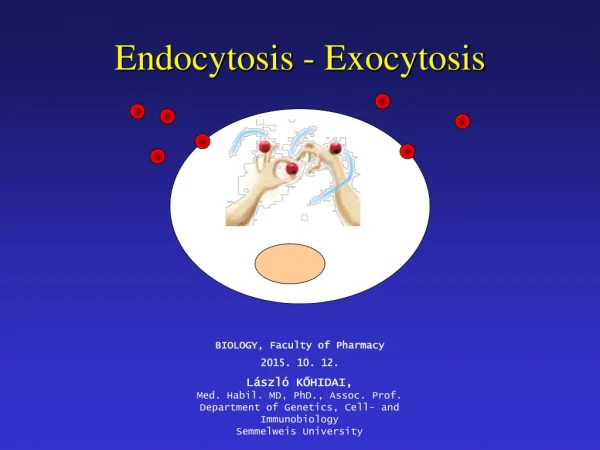
Endocytosis and Exocytosis Advanced laboratory techniques Third stage Lecture (8) ( Theortical) Asisstant lecturer RajaaSaihood Abbas
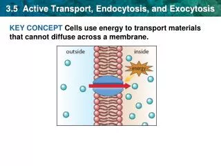
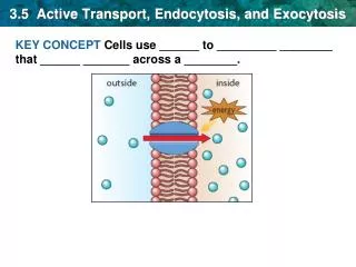
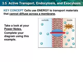
Endocytosis and Exocytosis • Exocytosis also known as 'reverse pino-cytosis' is the durable process by which a cell directs the contents of secretory vesicles out of the cell membrane. These membrane-bound vesicles contain soluble proteins to be secreted to the extracellular environment, as well as membrane proteins and lipids that are sent to become components of the cell membrane. • Endocytosis is an energy-using process by which cells absorb molecules (such as proteins) by engulfing them. It is used by all cells of the body because most substances important to them are large polar molecules that cannot pass through the hydrophobic plasma or cell membrane.
Types of endocytosis • 1. Pinocytosis. • 2. Receptor-Mediated Endocytosis. • 3. Phagocytosis.
Pinocytosis • 1.Pinocytosis ( cell drinking) Pinocytosis, is a process by which liquid droplets are ingested by living cells. Pinocytosis is one type of endocytosis, the general process by which cells engulf external substances, gathering them into special membrane-bound vesicles contained within the cell. In pinocytosis, rather than an individual droplet of liquid traveling passively through the cell membrane, the droplet first becomes bound, or adsorbed, on the cell membrane, which then invaginates (forms a pocket) and pinches off to form a vesicle in the cytoplasm.
Receptor- Mediated Endocytosis • 2.Receptor- Mediated Endocytosis • (also called clathrin-dependent endocytosis) Some of integral membrane proteins that a cell displays at its surface are receptor for particular components of the extracellular fluids (ECF) . • For example , iron is transported in the blood complex to a protein called transferrin . Cells have receptors for transferrin on their surface .when these receptors encounter a molecule of transferring, they bind tightly to it. The complex of transferrin and its receptor is then engulfed by endocytosis. • Ultimately, the iron is released into the cytosol. Receptor- Mediated endocytosis is many thousand times more efficient than simple pinocytosis in enabling the cell to acquire the macromolecules it needs.
Phagocytosis • 3.Phagocytosis: • Polymorphonuclearneutrophilic leukocytes have been well-known components of the innate immune system for many years. Detailed studies of PMN phagocytosis and intracellular killing of microorganisms have led to a better understanding of important defense mechanisms against invasion by pathogenic bacteria, fungi, and enveloped viruses. PMNs are attracted to the site of microbial invasion, recognize the microbe, become activated, kill the microorganisms, resolve the infection, undergo apoptosis, and are then ingested and removed by either macrophages or neighboring endothelial cells to resolve the inflammatory response. Phagocytosis is a form of endocytosis. In the process of phagocytosis the cell changes shape by sending out projections which are called pseudopodia (false feet).
Phagocytic cell • The phagocytic cell such as a macrophage may be attracted to a particle like a bacteria or virus by chemical attractant. This process is called Chemotaxis (movement toward a source of chemical attractant). • Activation of phagocytosis causes changes in the cytoskeletal contractile elements, which leads to an invagination of the cell membrane of the PMNs. This occurs at the site of the attachment of the opsonized microorganism. Pseudopods extend from the PMNs and fuse around the invagination encasing the microorganism inside the phagolysosomal vacuole.
Some sort of receptor ligand interaction occurs between the phagocytic cell surface and the particle that will be ingested. The pseudopodia then surround the particle and when the plasma membrane of the projection meet membrane fusion occurs. This results in the formation of an intracellular vesicle.
Phagocytosis • The process of phagocytosis begins with attachment and ingestion of microbial particles into organelle called phagosome. Once inside the phagocyte, the phagosome containing the microorganism joins with a lysosome which contributes enzymes .The fusion of phagosome and lysosome result in phagolysosome, microorganisms are destroyed within minutes. Chemical portions of microorganism called antigenic determinants are displayed on the surface of the phagocyte to stimulate the immune process.
Phagocyte • Phagocyte A cell, such as a white blood cell, that engulfs and absorbs waste material, harmful microorganisms, or other foreign bodies in the bloodstream and tissues. Several types of cells in the immune system engulf microorganisms via Phagocytosis: • Neutrophils. Neutrophils are abundant in the blood, quickly enter tissues, and phagocytize pathogens in acute inflammation. • Macrophages. Macrophages are closely related to monocytes in the blood. These longer-lived cells predominate in chronic inflammation.. • Dendritic Cells. • B Lymphocytes. A small amount of phagocytosis in these cells is often necessary in order for them to develop into cells that release antibodies.
In human and in vertebrates generally the most effective phagocytic cells are two kinds of leukocytes 1- the Macrophage (large phagocytic cells) and 2- the Neutrophils. • The macrophage occur especially in the lungs, liver, spleen and lymph nodes, where their function is to free the airways, blood and lymph from bacteria and other particles. Macrophages also are found in all tissues as wandering amoeboid cells, and the monocyte. • The smaller phagocytes are chiefly neutrophils that are carried along by the circulating blood until they reach an area of infected tissue , where they pass through the blood vessel wall and lodge in that tissue. Both macrophages and neutrophils are drawn toward an area of infection or inflammation by means of substances given off by bacteria and the infected tissue or by a chemical interaction between the bacteria and the complement system of blood serum proteins.
PHAGOTEST • PHAGOTEST • (Test Kit For The QUANTIFICATION OF PHAGOCYTIC ACTIVITY • OF MONOCYTES AND GRANULOCYTES IN HEPARINIZED WHOLE BLOOD) • This test kit allows the quantitative determination of leukocyte phagocytosis in heparinized whole blood. It contains fluorescein (FITC)-labelled opsonized bacteria (E. coli-FITC) and necessary reagents. It measures • the overall percentage of monocytes and granulocytes showing phagocytosis in general (ingestion of one or more bacteria per cell) and the individual cellular phagocytic activity (number of bacteria per cell).
PHAGOTEST • The investigation of phagocytosis can be performed either by flow cytometry or by fluorescence microscopy. • PHAGOTEST : is intended to investigate the phagocytic activity found in various disorders and to evaluate the effects of drugs. • Abnormal phagocytosis can occur with a variety of disorders. The defects can be associated with the neutrophil itself or with an immunoglobulin or complement defect. Acquired defects associated with altered phagocytic activity can be observed in trauma, diabetes, renal failure, and infection. Reduced phagocytosis has been observed in recurrent bacterial skin and pulmonary infections, in wound infections from burns , in patients with AIDS. • Various immunomodulators (cytokines such as interleukin-2 or interferon-1 , lactic acid bacteria and plant extracts ) can increase the phagocytic activity of neutrophils and monocytes. These effects can be investigated in vitro or ex vivo .