Download
1 / 22
220 likes | 337 Vues
Microscopes—tools for studying biodiversity. detect, distinguish between organisms study structure of cells, tissues, organs determine locations of molecules. simple microscope. 1 lens (eg magnifying glass) bends light so object appears larger. compound microscope. >1 lens mag multiplies

E N D
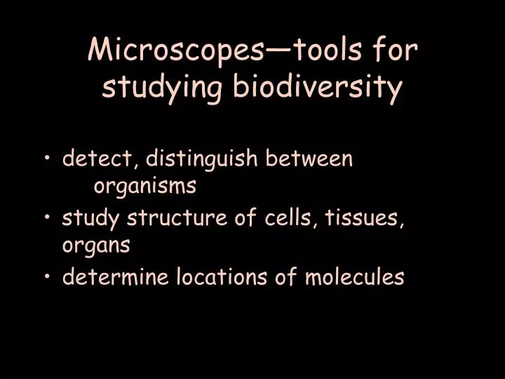
Microscopes—tools for studying biodiversity • detect, distinguish between organisms • study structure of cells, tissues, organs • determine locations of molecules
simple microscope • 1 lens (eg magnifying glass) • bends light so object appears larger
compound microscope • >1 lens • mag multiplies • so do optical problems
optical terms • magnification: how large appears compared to actual size • resolution (resolving power): closest distance 2 objects can be, & still be distinguished as separate obj. • resol dep on wavelength () • shorter better. electron microscopes
lens resolution comparison • human eye 0.2mm ruler in lab • light scope 0.2m 1000x better than eye • electron scope ~ 2 nm 100x better than LM 100,000x better than eye • = micro • LM = light scope • EM = electron scope
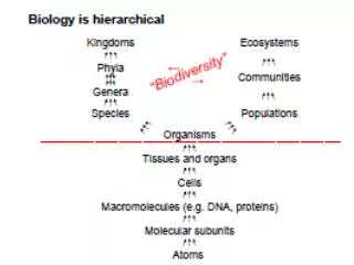
scale • 1mm = 1000 m = 1 million nm • human hair ~1/10 of mm = 100 m • electron microscopy reveals viruses! 20 – 90 nm • 1990s: 50 million/mL of seawater, soil • Fig 6.2, p 95
image terms • photograph--no scope • photomicrograph--photo taken though scope • light micrograph • electron micrograph
LM • Advantage: living cells & organisms • colors (pigments) • movement • focus through depth of specimen
EM • disadvantage: specimens dead • electrons from filament • in vacuum • focus with electromagnetic lenses
TEMtransmission electron microscope • electrons go through specimen • make shadow • fine structure inside of cells • many sections for 3-D structure
TEM • can locate molecule w/antibody attached to gold particle
SEM scanning electron microscope • electron beam scans specimen • specimen electrons collected • surface of object • 3-D view
LM contrast • pigments • stains • brightfield--standard bright background • special techniques (Fig 6.3, p 96) • polarized light • darkfield • differential interference contrast (dic) • phase contrast
fluorescence microscopy • UV light source • specimen emits light • autofluorescence • tagged antibodies • reporter proteins
Confocal microscopy • laser light source • optical sections • no out of focus blur
discovery of cells • 1665 English scientist Robert Hooke • first saw (cork) cells [cell = room] • compound microscope • (30x mag) • Royal Society, London Hooke’s drawing of cork cells in Micrographia http://www.gutenberg.org/files/15491/15491-h/15491-h.htm section 18, plate 11
Hooke’scompound microscope http://www.arsmachina.com/hooke.htm
scientific literature • primary lit. — original report of research • intro, materials & methods, results, discussion • peer-reviewed • secondary lit. — report about primary lit. • ProcRSocL, Nature, PNAS, Science
discovery of microorganisms • 1674 Dutch merchant Antony van Leeuwenhook • first saw “animalcules” • protists, and later bacteria • simple microscope w/great lens • 275-295x mag http://www.ucmp.berkeley.edu/history/leeuwenhoek.html
discovery of microorganisms • 1676 Leeuwenhook wrote to Royal Society @ singled celled organisms • Hooke confirmed • first published bacteria • voucher specimens http://www.ucmp.berkeley.edu/history/leeuwenhoek.html
Leeuwenhoek’s microscope light micrograph of red blood cells photographed through this scope http://www.brianjford.com/wav-mict.htm