Download

1 / 18
200 likes | 288 Vues
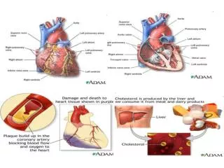
BLOOD SUPPLY OF THE HEART. Dr Jamila EL medany. &. Dr Essam Salama. Objectives. At the end of the lecture the student should be able to know about; The arterial supply of the cardiac muscle regarding (origin, course, distribution and branches). The coronary anastmosis.

E N D
BLOOD SUPPLY OF THE HEART Dr Jamila EL medany & Dr Essam Salama
Objectives • At the end of the lecture the student should be able to know about; • The arterial supply of the cardiac muscle regarding (origin, course, distribution and branches). • The coronary anastmosis. • The arterial supply to the conducting system of the heart. • The venous drainage of the heart regarding (origin, tributaries and termination).
Arterial Supply The arterial supply of the heart is provided by TwoCoronary Arteries : Right Coronary artery & Left Coronary artery They are distributed over the cardiac surface, within the subepicadium connective tissue. L Coronary artery
Origin of Coronary Arteries LCA RCA • They arise fromthe initial part of theAscending Aorta (Aortic Sinuses), immediately above the aortic valve. AA
Right Coronary Artery Arises from the anterior aortic sinus of the ascending aorta. Descends in the right atrioventricular groove between the Right Auricle and the Pulmonary trunk. At the inferior border of the heart it continues posteriorly to anastomose with the left coronary.
(RCA ) Supplies: • Right atrium, • Right ventricle, • part of Left Atrium, • Left ventricle & Atrioventricular septum. • Most of conducting system
Branches Right Conus; For infundibulum and upper part of anterior wall of the right ventricle. Anterior ventricular branches; 2-3 branches supply anterior surface of the right ventricle. Marginal arteryis the largest branch, runs along the lower margin of the sternocostal surface. It is accompanied by the Small Cardiac vein. Right conus
Atrial branches; Supply anterior and lateral surfaces of the right atrium One branch supplies posterior surface of both atria Artery of the Sinuatrial Node Supplies the SAN and both atria In 35% it arises from the left coronary. Posterior ventricular branches; About 2 supply the diaphragmatic surface of the right ventricle. Posterior Interventricular artery;(accompanied by Middle Cardiac vein) Lies in the posterior interventricular groove, it supplies the Right and Left Ventricles, including their inferior wall, posterior part of ventricular septum, Not the Apical part,
Left Coronary Artery The Larger of the two coronaries. Arises from theleft posterior aortic sinusof the ascending aorta. Descends: 1. Between the pulmonary trunk and the left auricle. 2. In the IV groove to the apex of the heart. Divides into two terminal branches: Anterior Interventricular & Circumflexarteries. .
Branches (A)Anterior Interventricular Descends in the anterior interventricular groove to the apex of the heart (accompanied by the Great cardiac vein) in most individuals it passes around the apex to anastomose with terminal branches of the right coronary , in 1\3 it ends at the apex ) It supplies the right and left ventricles and anterior part of ventricular septum It gives: 1. Left conus artery for pulmonary conus. 2. Anterior ventricular and Posterior ventricular ; Supply left ventricle 3. Atrial branches ; Supply greater part of left atrium
4. Left diagonal artery • one of the ventricular branches or may arises from left coronary, • (B) Circumflex Artery; • Winds around the left margin of the heart in the atrioventricular groove • C. Left Marginal artery ; • Supplies the left margin of the left ventricle down to the apex
Variations of the Coronary Arteries • Right Dominance: • In (90 %) of population, the Posterior Interventricular artery is a branch of the Right Coronary. • Left Dominance: • In the rest (10%), the Posterior Interventricular artery arises from the Circumflex branch of the Left Coronary A
Coronary Anastomosis • In MOST of people, the terminal branches of the right and left coronaries anastomose in the posterior part of the IV groove • However this anastomoses is not large enough to provide adequate blood supply in case of coronary occlusion, (Functional End arteries)
Arterial Supply ofConducting System • SAN, AVN & AVB are usually supplied by Right coronary. • Right Bundle Branch (RBB) of (AVB) is supplied by Left coronary • LBB of (AVB) is supplied by both Right and Left coronaries
Coronary Sinus • Drains most of the Venous Blood of the heart. • It lies in the Posterior part of the AV groove. • Origin : • It is the direct continuation of the Great Cardiac Vein. • Tributaries: • (3) Cardiac Veins: • A. Great. • B. Middle. • C. Small. • Oblique vein of left atrium (vein of Marshall).
Termination: • It empties into Right Atrium. • Its opening is inferior & to the left of the IVC opening. • It is guarded by a valve.
Veins Draining outside Coronary Sinus • 1. Anterior cardiac veins: • Open directly into the Right Atrium. • 2. Venae Cordisminime (small cardiac veins): • Open into the heart chambers.