Download
1 / 7

E N D
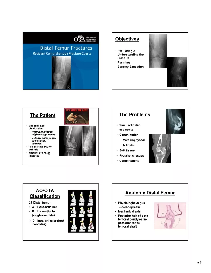
Objectives Orthopaedic Trauma Association Distal Femur Fractures Resident Comprehensive Fracture Course • Evaluating & Understanding the Fracture • Planning • Surgery Execution Protection and/or confidentiality of contents statement, this statement may also include a corporate copyright notice. The Problems The Patient • Small articular segments • Bimodal age distribution – young healthy pt, high energy, males – elderly, osteopenic, low energy, females • Pre-existing injury/ arthritis • Amount of energy imparted • Comminution – Metadiaphyseal – Articular • Soft tissue • Prosthetic issues • Combinations AO/OTA Classification Anatomy Distal Femur •A1 A2 A3 33 Distal femur • A Extra-articular • B Intra-articular (single condyle) • Physiologic valgus – (5-9 degrees) • Mechanical axis • Posterior half of both femoral condyles lie posterior to the femoral shaft •B1 B2 B3 C Intra-articular (both condyles) •C1 C2 C3 • 1
Anatomy Distal Femur What This Means for Fixation • Femur transitions from cylinder to condyles • Medial condyle extends further inferior • Cancellous bone • Trapezoidal shape Avoid notch and concomitant injury to cruciate ligaments •X Avoid penetration of medial cortex with anterior screws •X Radiographic Exam Radiographs AP/lateral knee & femur AP/lateral contralateral distal femur for planning CT scan Joint details Coronal split Sagittal split Deforming forces • Quadriceps, hamstrings shorten • Gastrocnemius extends at fx, rotation of intercondylar split • Other forces from cruciates, capsule, popliteus, collateral ligaments External Fixation? Spanning knee external fixation – Allows for temporary stabilization of fracture if delayed reconstruction is necessary – External fixator as a reduction aid at time of definitive reconstruction Keep pins out of planned surgical field! Planning • 2
Plan Ahead Internal Fixation Options Principles of surgical treatment: 1. Careful handling of soft tissues 2. Anatomic reduction of the articular surface and restoration of limb axial alignment, rotation, and length 3. Indirect reduction techniques 4. Stable internal fixation 5. Early rehabilitation Condylar buttress plates Fixed-angle devices – Blade plate – Dynamic Condylar Screw (“DCS”) Retrograde intramedullary nail Locked plates All implants can work if utilized properly! Plating Plan of Attack • Advantages – May be able to get fragment specific screws through plate – Better than nail for significant trochlea comminution • Disadvantages – Blood loss? – Medial Hoffa fragment difficult to see through anterolateral incision – Does not reduce the fracture (but can fine tune reduction) 1) Reduce articular surfaces first – Direct reduction techniques 2) Secure fixation of articular surfaces – Interfragmentary screws – Must consider where other hardware will go (e.g. locking screws) 3) Restore continuity of articular block with shaft – Indirect reduction techniques Reduction Reduction • Reduce the Hoffa • Restore the articular surface • Reduce the metaphysis to to the diaphysis • Indirect reduction aids – Bump – Ex fix • Check your lateral for alignment and plate position proximally •Tip: Notice K-wires driven thru medially and out of way for plate • 3
Reduction • Reduction completed before plate applied • Check reduction as plate is applied on both sides of fracture • Stiffness determined by plate material properties and screw spread around fracture Reduction • First screws distally • Then secure proximally – Ensure plate in good position Indirect Reduction Indirect Reduction Indirect reduction techniques – External fixator – Femoral distractor – “Joysticks” – Percutaneous clamps – Bumps – Blocking screws Not for articular surfaces – Direct visualization and reduction Preserves soft-tissue envelope around metadiaphyseal fracture lines – Achieve restoration of length, alignment, and rotation via traction and manipulation utilizing reduction aids that do not strip soft tissues around the fracture site Respect the Biology – Indirect Reduction Limit soft tissue dissection – Indirect reduction techniques – Submuscular plate application without extensive stripping – Preserve periosteal blood supply when able • 4
• Don’t forget to bone graft if necessary • Consider intramedullary implant if no medial support Retrograde IMN Retrograde Nailing Retrograde Nailing • Clinical Advantages – Implant failure less likely to be catastrophic – Implant placement not as technically challenging • Can avoid translational and varus/valgus deformity – Better access to Hoffa fractures through anterior arthrotomy (medial or lateral) – Generally faster and less invasive – Protects entire bone – Load sharing: earlier weightbearing? • Disadvantages – Need access to entire intramedullary canal – More difficult with trochlear comminution • Compromised start point Retrograde Nailing Retrograde Nailing-Beware! • Biomechanical Advantages – Shorter moment arm for interlocks compared to locking screws in lateral plate – Improved axial stability compared to plate – More distal fixation (nail plus locked bolt) •Can be difficult for complex distal femur fractures – know your limitations! • 5
Extraarticular Distal Femur Caution!! Retrograde IMN Don’t forget to reduce the fracture before placing distal interlocks! – Nail will not have an isthmic fit – May need blocking pins/drill bits/screws – Percutaneous or open clamps – Nail will “lock” a fracture in a malreduced position as easily as it will “lock” a fracture reduced • Most Common Deformity is Apex Posterior – Adjust position of bump – Small lateral incision and clamp fracture before locking – May require blocking drill bits/pins/screws Blocking Pins, Drill Bits, and Screws Retrograde IMN Theoretical “Disadvantages” – Arthrotomy required • Is this inherently bad? • Also generally performed for locking distal femur plates – “Percutaneous” joint fixation • Just because you use a nail doesn’t mean you can’t expose the joint – Lack of alignment control (“windshield wipering” of implant • Not an issue is nail is used appropriately – Difficulty of insertion with TKA • Make an arthrotomy and look right at your starting point! • Using the nail as a reduction tool to correct angular deformities • Does not substitute for a good start point • 6
Avoiding Errors in Judgement • Make a Problem List –Soft Tissue considerations –Hoffa? –Articular Reduction –Restoring Metadiaphyseal relationship –Controlling Stiffness of Implant Summary Make A Plan • Approach • Plate or nail? • Fragment-specific screws • Reduction Aids Questions? • 7