1 / 74

Chapter 9 Opener

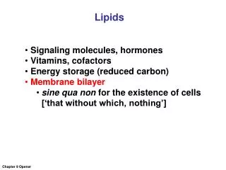
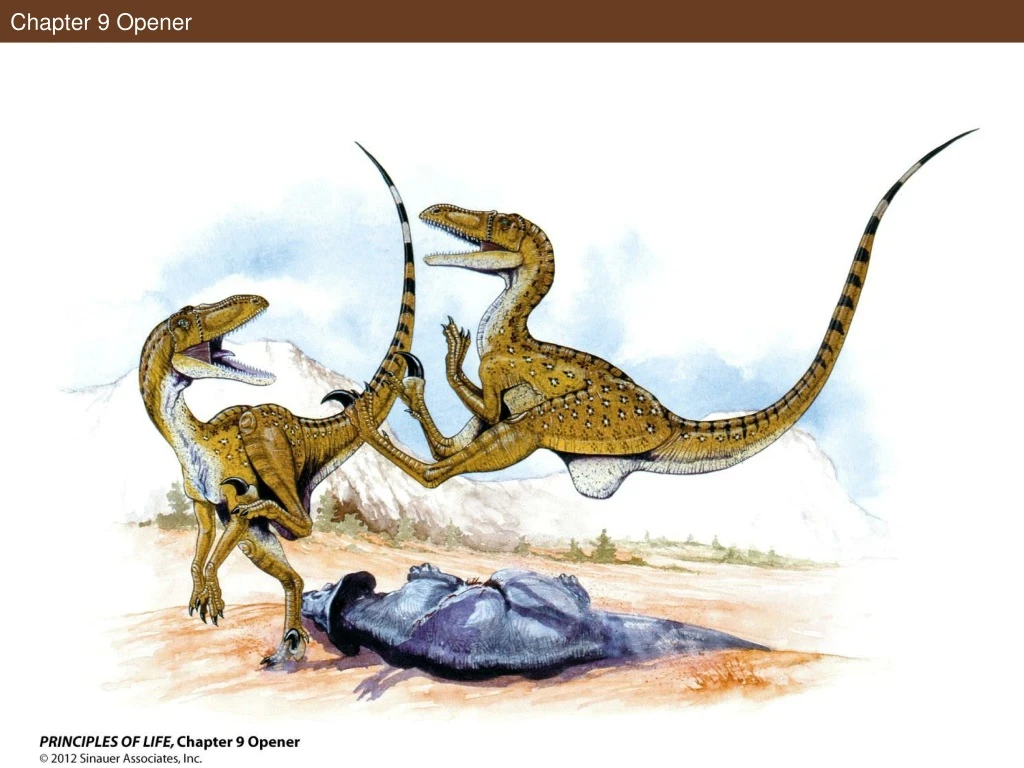
Chapter 9 Opener. Figure 9.1 DNA in the Nucleus and in the Cell Cycle. Figure 9.1 DNA in the Nucleus and in the Cell Cycle. Figure 9.1 DNA in the Nucleus and in the Cell Cycle (Part 1). Figure 9.1 DNA in the Nucleus and in the Cell Cycle (Part 2). In-Text Art, Ch. 9, p. 166.

mmayfield
Télécharger la présentation

Chapter 9 Opener

An Image/Link below is provided (as is) to download presentation Download Policy: Content on the Website is provided to you AS IS for your information and personal use and may not be sold / licensed / shared on other websites without getting consent from its author. Content is provided to you AS IS for your information and personal use only. Download presentation by click this link. While downloading, if for some reason you are not able to download a presentation, the publisher may have deleted the file from their server. During download, if you can't get a presentation, the file might be deleted by the publisher.

E N D

Presentation Transcript


  1. Chapter 9 Opener

  2. Figure 9.1 DNA in the Nucleus and in the Cell Cycle

  3. Figure 9.1 DNA in the Nucleus and in the Cell Cycle

  4. Figure 9.1 DNA in the Nucleus and in the Cell Cycle (Part 1)

  5. Figure 9.1 DNA in the Nucleus and in the Cell Cycle (Part 2)

  6. In-Text Art, Ch. 9, p. 166

  7. Figure 9.2 Viral DNA and Not Protein Enters Host Cells

  8. Figure 9.2 Viral DNA and Not Protein Enters Host Cells

  9. Figure 9.2 Viral DNA and Not Protein Enters Host Cells (Part 1)

  10. Figure 9.2 Viral DNA and Not Protein Enters Host Cells (Part 2)

  11. Figure 9.3 Transformation of Eukaryotic Cells

  12. Figure 9.3 Transformation of Eukaryotic Cells

  13. Figure 9.3 Transformation of Eukaryotic Cells (Part 1)

  14. Figure 9.3 Transformation of Eukaryotic Cells (Part 2)

  15. Figure 9.4 X-Ray Crystallography Helped Reveal the Structure of DNA

  16. Figure 9.4 X-Ray Crystallography Helped Reveal the Structure of DNA

  17. Figure 9.4 X-Ray Crystallography Helped Reveal the Structure of DNA (Part 1)

  18. Figure 9.4 X-Ray Crystallography Helped Reveal the Structure of DNA (Part 2)

  19. In-Text Art, Ch. 9, p. 169

  20. In-Text Art, Ch. 9, p. 169

  21. Figure 9.5 DNA Is a Double Helix

  22. Figure 9.5 DNA Is a Double Helix

  23. Figure 9.5 DNA Is a Double Helix (Part 1)

  24. Figure 9.5 DNA Is a Double Helix (Part 2)

  25. Figure 9.6 Base Pairs in DNA Can Interact with Other Molecules

  26. Figure 9.6 Base Pairs in DNA Can Interact with Other Molecules

  27. In-Text Art, Ch. 9, p. 172

  28. Figure 9.7 Each New DNA Strand Grows by the Addition of Nucleotides to Its 3′ End

  29. Figure 9.7 Each New DNA Strand Grows by the Addition of Nucleotides to Its 3′ End

  30. Figure 9.8 The Origin of DNA Replication

  31. Figure 9.8 The Origin of DNA Replication

  32. Figure 9.8 The Origin of DNA Replication (Part 1)

  33. Figure 9.8 The Origin of DNA Replication (Part 2)

  34. Figure 9.9 DNA Forms with a Primer

  35. Figure 9.9 DNA Forms with a Primer

  36. Figure 9.10 DNA Polymerase Binds to the Template Strand

  37. Figure 9.10 DNA Polymerase Binds to the Template Strand (Part 1)

  38. Figure 9.10 DNA Polymerase Binds to the Template Strand (Part 2)

  39. Apply the Concept, Ch. 9, p. 175

  40. Figure 9.11 The Two New Strands Form in Different Ways

  41. Figure 9.11 The Two New Strands Form in Different Ways

  42. Figure 9.12 The Lagging Strand Story

  43. Figure 9.12 The Lagging Strand Story

  44. Figure 9.12 The Lagging Strand Story (Part 1)

  45. Figure 9.12 The Lagging Strand Story (Part 2)

  46. Figure 9.12 The Lagging Strand Story (Part 3)

  47. Figure 9.13 Telomeres and Telomerase

  48. Figure 9.13 Telomeres and Telomerase

  49. Figure 9.13 Telomeres and Telomerase (Part 1)

  50. Figure 9.13 Telomeres and Telomerase (Part 2)

More Related