Download
1 / 55
560 likes | 605 Vues
Common Ophthalmic Conditions in Primary Care - Management of Dry Eyes. Mr Yajati K Ghosh FRCSEd Consultant Ophthalmic Surgeon Birmingham & Midland Eye Centre. Affiliations. BMI Droitwich BMI Priory , Edgbaston Spire Parkway, Solihull. Subspeciality interests. Lid and lacrimal surgery

E N D
Common Ophthalmic Conditions in Primary Care - Management of Dry Eyes Mr Yajati K GhoshFRCSEd Consultant Ophthalmic Surgeon Birmingham & Midland Eye Centre
Affiliations • BMI Droitwich • BMI Priory, Edgbaston • Spire Parkway, Solihull
Subspeciality interests • Lid and lacrimal surgery • Periocular cancer surgery • Phacoemulsification Cataract surgery • Management of watery eyes
Common Ophthalmic Conditions Red Eye • Blepharitis • Conjunctivitis • Dry eyes • Watery eyes • Foreign body • Keratitis • Uveitis • Lumps & Bumps • Cataracts
What to do? 50% of ALL red eyes and irritable eyes are due to Dry Eye related conditions ………..
Overview http://www.virtualcancercentre.com
Roles and Characteristics of the eyelids • Eye protection • Regular blink: protection and stability of the tear film • Rich of glands • Adequate blood supply
The Anatomy • Corneal epithelium • Conjunctival epithelium • Tear film • Clinical ocular surface consist of conjunctiva cornea eyelids lacrimal gland lacrimal passages
www.virtualmedicalcentre.com Tear Secretion • Lacrimal gland Producing the watery part of the tear film called the aqueous. • Meibomian glands Producing lipids which keep the tear film from evaporating. • Goblet cells of the conjunctiva Producing mucin which allows the wetting of the ocular surface as well as stabilizes the tear film.
Tear and the Tear Film • Function : 1.Cleaning 2.Wetting ocular surface 3.Bacteriostasis 4.Supporting the cornea (oxygen supply) http://www.drmalcolmmckellar.co.nz
Concept • The ocular surface is a complex biological continuum responsible for the maintenance of corneal clarity, elaboration of a stable tear film for clear vision, as well as protection of the eye against microbial and mechanical insults. • Comprising a variety of disorders on cornea, eyelid, conjunctiva, lacrimal apparatus and tear film.
Healthy tear film Dry eye Dry Eye http://www.chronicdryeye.com
http://www.drmalcolmmckellar.co.nz Etiological factor & Classification • Aqueous tear deficiency • Lipid tear deficiency • Mucoprotein deficiency • Kinetic disorders of lacrimal fluid
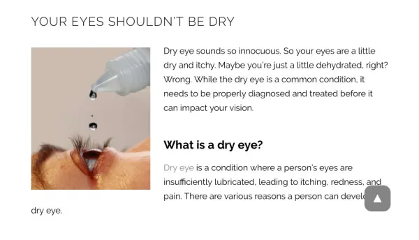
Conception • Dry eye - is a chronic lack of sufficient lubrication and moisture in the eye. • Its consequences range from subtle but constant irritation to ocular inflammation of the anterior (front) tissues of the eye.
Clinical Manifestation • Dry eye symptoms asthenopia irritation, grittiness dryness burning light sensitivity pink-eye • Do you regularly experience one or several symptoms above? • Some diseases and conditions (like Rheumatoid arthritis, Lupus and Sjögren’s Syndrome) also cause chronic Dry Eye in many patients. • Activities like Reading, Wearing contact lenses or Working on the Computer may cause Dry Eye.
Diagnostic Tests for Dry Eye • Dry Eye questionnaire • Lacrimal river width • Schirmer test – uses paper strips under eyelid to measure the wetness that collects over a specific period of time. • Break-up time of tear film (BUT) • Staining – uses special dyes to highlight areas of possible damage to the eye surface. • Tear lab – measuring tear osmolarity • Tear ferning test • Lactoferrin contents • Tear penetration pressure test • Corneal tonographic map • Impression cytology
Diagnosing • Schirmer test, BUT, Staining • Foundation Symptom Instability of tear film Damage to epithelium Tear penetration pressure increasing
SchirmerTest • Normal:≥10mm/5min
0分 1分 3分 2分 Staining • Using special dyes to highlight areas of possible damage to the eye surface.
http://www.dryeyezone.com Meibomian Gland Dysfunction
http://www.revophth.com Etiological Factor • Failure of the glands to produce or secrete lipids. • Wax ester declining and cholesterol increasingmake the symptoms worse . • Lack of tears and tear penetration pressure increasing. • Lupus, brandy nose etc.
Figure: Notching of the lid caused by loss of meibomian glands. http://www.eyehealthnutrition.com Clinical Manifestation • Common in aged people and who livedin cold region. • No specific symptoms. • Lid-margin mostly thickening; abnormal secretion while pressurizing. • Disorder in Meibomian gland, eyelid, conjunctiva.
Figure: No visible meibomian gland orifices: Eversion of the lower lids in both eyes showed atresic meibomian glands. http://www.ophmanagement.com Diagnosing • Absence of Meibomian gland. • The gland orifices are often compromised due to stenosis or closure. • A declining quality and quantity of lipid secretion. Any one of the physical signs can make the diagnosis of Meibomian gland dysfunction if the patient has clinical symptoms.
Treatment Clearing • Hot fomentation on eyelids for 5~10mins. • Massaging the eyelids. • Swabbing the lid-margin with mild cleaning solution.
Treatment • Antibiotics oral administration. • Local Medication Antibiotic eye drops Glucocorticoid eye drops (short term) Artificial tears
Classification • Corneal, conjunctival lesion Squamous epithelization type Limbal stem cell deficiency type • Tear film disorders Aqueous tear deficiency Lipid tear deficiency Mucoprotein deficiency Kinetic disorders of lacrimal fluid
Treatment • Reconstruction Epithelium, limbal stem cells Lacrimal secretion, tear film Innervation (nerve restore) Structure and function of eyelid • Surgical operation To re-establish conjunctiva, cornea, tear film and eyelid.
Treatment • According to the clinical category For tear deficiency: Maintain moisture in the eyes; reducing the evaporation; increasing the secretion; controlling inflammation & immunoreaction. For excess-evaporation: Therapy of Meibomian gland dysfunction; controlling inflammation; cleaning eyelid; decreasing the evaporation; lipid replacement. • According to the eye conditions For intermittent symptoms: Artificial tears add volume to the tear film as long as they remain in contact with the surface of the eye. For midrange dry eye: Artificial tears and punctal occlusion. For Severe dry eye: Appending cyclosporin, surgery.
Literature Artificial tears potpourri: a literature review Majid Moshirfar,1Kasey Pierson,2,*Kamalani Hanamaikai,3,*Luis Santiago-Caban,1Valliammai Muthappan,1 andSamuel F Passi1 Clin Ophthalmol. 2014; 8: 1419–1433. Published online 2014 Jul 31. doi: 10.2147/OPTH.S65263 PMCID: PMC4124072
Artificial Tear Groups HPMC Carboxy Methyl Cellulose PVA Liquid Polyols Hyaluronic acid Inserts Miscellaneous