Download
1 / 60
610 likes | 873 Vues
ASSESSMENT OF NEUROLOGICAL FUNCTION MICHELLE GARDNER RN, MSN. OBJECTIVES. Review the structures and functions of the central and peripheral nervous systems Describe the significance of physical assessment to the diagnosis of neurologic dysfunction.

E N D
OBJECTIVES • Review the structures and functions of the central and peripheral nervous systems • Describe the significance of physical assessment to the diagnosis of neurologic dysfunction. • Describe diagnostic tests used for assessment of suspected neurologic disorders and related nursing implications • Describe the needs of patients with various neurologic dysfunctions
NEUROLOGIC OVERVIEW • Central nervous system (CNS) - brain and spinal cord • Peripheral nervous system - cranial/spinal nerves - autonomic nervous system • Basic functional unit neuron
Function of the Nervous System • Control all motor, sensory, autonomic, cognitive, and behavioral activities
Central Nervous System • The Brain cerebrum brain stem cerebellum
Peripheral Nervous System Include • Cranial nerves • Spinal nerves • Autonomic nervous system
Autonomic Nervous System (ANS) • Functions to regulate activities of internal organs and to maintain and restore internal homeostasis. • Sympathetic NS - “fight or flight responses • Parasympathetic NS - controls most visceral functions - serves to conserve and restore the energy stores in the body
Neurological Assessment Health history • History of the present illness-DETAILS • Review the medical records • Input from witness/family member
Neurological Assessment Common symptoms • Pain • Seizures • Dizziness/vertigo • Visual disturbances • Muscle weakness • Abnormal sensations
Diagnostic Evaluation • CT scan (Computer Tomography) • MRI (Magnetic Resonance Imaging) • PET (Positron Emission Tomography) • Cerebral angiography • Electroencephalography (EEG) • Electromyography (EMG) • Lumbar puncture – analysis of CSF
CT Scan • Computer – assisted x-ray of multiple cross sections of the brain to detect problems hemorrhage, brain atrophy, infection, tumor and other abnormalities. • Contrast media may be used • Assess for contraindications to contrast media shell fish/iodine/dye allergy • Explain appearance of scanner • Instruct client to remain still during the procedure. • Evaluate renal function
Magnetic Resonance Imaging (MRI) • Imaging of brain, spinal cord by means of magnetic energy. • Used to detect strokes, tumors, seizures, trauma • Not an invasive procedure • Has greater contrast in images of soft tissue structures than CT scan. • Contrast media may be used to enhance images. • Screen client for metal parts
Electroencephalography -EEG • Electrical activity of the brain is recorded by scalp electrodes to evaluate seizure disorders, cerebral diseases, brain death. • Procedure is noninvasive and without danger of electrical shock. • Medication may be withheld • Resume medication and wash electrode paste out of hair after the test.
Cerebral Angiography • X-ray visualization of intracranial/extracranial blood vessels viewed to detect vascular lesions and tumors of the brain. • Contrast medium is used/explain procedure. • Assess client for stroke risk before procedure • Monitor neurological signs and VS • Report any neurological changes
Electromyography EMG • Electrical activity associated with nerve and skeletal muscle is recorded by insertion of needle electrodes to detect muscle and peripheral nerve disease. • Inform client that pain and discomfort may be associated with procedure insertion of needles.
Lumber Puncture Cerebrospinal fluid analysis • CSF is aspirated by needle insertion in L3-4 or L4-5 interspace to assess many CNS diseases • Client assumes and maintains lateral recumbent position • Ensure strict aseptic technique • Post procedure- headache • CONTRAINDICATED with patients with ICP
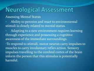
Consciousness • Person is aware of self and the environment and is able to respond appropriately to stimuli • Full consciousness requires both alertness and full cognition
Altered LOC - • Altered LOC is not a disorder but the result of a pathology • Full consciousness • Confusion • Disorientation • Obtundation • Coma
Pathophysiology A-E-I-O-U = • Alcohol, Epilepsy, Insulin, Opium, Uremia TIPSS = • Tumor, Injury, Psychiatric, Stroke, Sepsis
LOC – Assessment • Assess verbal response and orientation • Alertness • Motor responses • Respiratory status • Eye signs • Reflexes • Posturing • Glasgow Coma Scale • Client is at risk for alterations in every body system
POSTURING Decorticate Posturing Decerebrate Posturing
Interdisciplinary Care • Must begin immediately Focus • identify the underlying cause • preserve function • prevent deterioration
Diagnostic Procedures • CT scan/MRI • EEG • Cerebral angiography • Laboratory tests - blood glucose - electrolytes - ABG - liver function test - toxicology screening
Potential Complications • Respiratory distress or failure • Pneumonia • Aspiration • Pressure ulcer • Deep vein thrombosis (DVT) • Contractures
Ineffective Airway Clearance • Assess/monitor • Positioning to prevent obstruction of upper airway—HOB elevated 30° • Suctioning, and CPT • Monitor ABG analysis
Impaired Physical Mobility • Frequent turning; use turning schedule • Passive ROM • Use of splints, foam boots, trochanter rolls, and specialty beds as needed • Clean eyes with cotton balls moistened with saline • Use artificial tears as prescribed
Risk for Imbalanced Nutrition - • Assess swallowing/gag reflex • Monitor and report manifestations of aspiration • Provide interventions to prevent aspiration • Monitor nutritional status • Assess the need for alternative methods of nutritional support - collaboration dietitian
Communication/Family Support • Encourage the family to talk to and touch patient • Maintain normal day/night pattern of activity • Orient the patient frequently • Note: When arousing from coma, a patient may experience a period of agitation; minimize stimulation at this time • Allow family to ventilate and provide support to them • Reinforce and provide consistent information to family • Referral to support groups and services for family
Increased Intracranial Pressure • Skull is like a closed box (3) essential volume components - brain tissue (80%) - blood (12%) - cerebrospinal fluid (8%) • These components equal a state of equilibrium and produce ICP. • ICP measured in the lateral ventricles normal pressure 10-15mmHg. 15mmHg being the upper limit.
Increased Intracranial Pressure Monroe-Kellie hypothesis • A state of equilibrium exist: if the volume of any of the three components increases, the volume of the others must decrease to maintain normal pressures within the cranial cavity . • Brain tissue has limited space to expand, compensation is accomplished by - displacing/shifting CSF, - increasing the absorption/diminishing the producing CSF - decrease cerebral blood volume
Increased Intracranial Pressure • Sustained elevated pressure within the cranial cavity • Caused by – head trauma, tumors stroke hemorrhage infection *cerebral edema
Increased Intracranial Pressure • Compensatory mechanism that compensate for increased ICP autoregulation and decreased production/flow of CSF . • Autoregulation – the brain’s ability to change the diameter of the blood vessels to maintain a constant cerebral blood flow.
Increased Intracranial Pressure ICP is increased by: • Endotracheal or oral tracheal suctioning • Coughing • Blowing nose forcefully • Head of bed less than 30 degrees • Increased intra-abdominal pressure(restrictive clothing, Valsalva)
Increased Intracranial Pressure Clinical Manifestations • Early sign – change in LOC • Motor responses • Vision & pupils • Vital signs • Other
Clinical Manifestations - late • Cushing’s triad: bradycardia, severe hypertension, bradypnea • projectile vomiting • further deterioration of LOCstupor to coma • decortication, decerebration • respiratory abnormalitiesCheyne-Stokes breathing • Headache
Increased Intracranial Pressure Diagnostic studies • CT scan/MRI • Serum Osmolality • ABG’s
Increased Intracranial Pressure Complications • Brain stem herniation • Diabetes inisipidus • Syndrome of inappropriate antidiuretic hormone (SIADH)