Download

1 / 83

E N D
Prosthetic Heart Valves Dr. Mona Youssef http://www.cardiologylinks.webs.com/home
complications • Primary valve failure • Prosthetic valve endocarditis (PVE) • Prosthetic valve thrombosis (PVT) • Thromboembolism • Mechanical hemolytic anemia • Anticoagulant-related hemorrhage
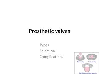
Make UP • Composition: - Synthetic material (mechanical prosthesis) - Biological tissue (bioprosthesis). - Homografts or preserved human aortic valves • Three main designs : - Caged ball valve - Tilting disc (single leaflet) valve - Bileaflet valve. *The only Food and Drug Administration (FDA)–approved caged ball valve is the Starr-Edwards valve
Bioprosthetic (xenograft) valves Made from: • Porcine valves e.g. Carpentier-Edwards valves or • Bovine pericardium e.g. Perimount series valves
Valve failure • Abruptly: -Tearing e.g. suture line dehiscence or breakage of components with acute valvular regurgitation or embolization of the valve fragments -Thrombus suddenly impinging on leaflet mobility • Gradually: - Calcifications with stiffening of the leaflets - Thrombus / pannus formation
Acute Mitral Valve Failure • Sudden left atrial volume overload • Increased left atrial pressure • Pulmonary venous congestion • Pulmonary edema • Decreased left ventricular output due to regurgitatnt volume into the left atrium • The compensatory mechanism of increased sympathetic tone: - Increases the heart rate and the systemic vascular resistance (SVR) - Decreases diastolic filling time - Impedes left ventricular outflow - Increases the regurgitant volume
Acute Aortic valve Failure • Rapidly progressive left ventricular volume overload • Increased left ventricular diastolic pressure • Pulmonary congestion and edema • Decreased cardiac output • The compensatory mechanism of increased sympathetic tone: - Increases heart rate and yields a positive inotropic state, aiding in the maintenance of cardiac output BUT also: -increases SVR impeding forward flow -Increases systolic wall tension yielding a rise in myocardial oxygen consumption& Myocardial ischemia, even in the absence of coronary artery disease.
Bioprosthesis • Mostly suffer from slow degeneration, calcification, or thrombus formation • Asymptomatic • gradually worsening congestive heart failure, with increasing dyspnea. • Unstable angina • Systemic embolization
Early PVEoccurring within 60 days of implantation Due to: perioperative contamination or hematogenous spread • Charecteristic lesion in mechanical valves : ring abscesses *Ring abscess may lead to: - Valve dehiscence and perivalvular leakage - Local extension with formation of myocardial abscesses - Further extension to the conduction system producing a new atrioventricular block - Less frequently valve stenosis and purulent pericarditis • Bioprosthetic valve PVE usually causes leaflet tears or perforations. Valve stenosis is more common with bioprosthetic valves than with mechanical valves. Ring abscess, purulent pericarditis, and myocardial abscesses are much less frequent in bioprosthetic valve PVE. • Finally: glomerulonephritis, mycotic aneurysms, systemic embolization, and metastatic abscesses
Late PVEoccurrs after 60 days usually caused by hematogenous spread
Incidence • Prosthetic valve thrombosis is more common in mechanical valves • Thromboembolic complication rates: - Hightest in Caged ball valves - Lowest in bileaflet valves • Valve thrombosis is increased with: - Valves in the mitral position - In patients with subtherapeutic anticoagulation. • Anticoagulant-related hemorrhagic complications : - Major hemorrhage in 1-3% of patients per year - Minor hemorrhage in 4-8% of patients per year.
hemolytic anemia - Low-grade in 70% of prosthetic heart valve recipients - Severe hemolytic anemia occurs in 3% *The incidence is increased with caged ball valves and in those with perivalvular leaks. • Primary valve failure occurs in: - 3-4% of patients with bioprostheses within 5 years of implantation and in up to 35% of patients within 15 years. - Mechanical valves have a much lower incidence of primary failure. • PVE occurs in 2-4% of patients: - 3% in the first postoperative year, then 0.5% for subsequent years. - The incidence is higher in mitral valves. - Mechanical and biological valves are equally susceptible.
Mortality/Morbidity • Acute failure of a prosthetic aortic valve usually leads to sudden or near-sudden death. • PVE has an overall mortality rate of 50%. • In early PVE, the mortality rate is 74%. • In late PVE, the mortality rate is 43%. • The mortality rate with a fungal etiology is 93%. • The mortality rate for staphylococcal infections is 86%. • Fatal anticoagulant-induced hemorrhage occurs in 0.5% of patients per year.
Age • The incidence of having any prosthetic valve complication decreases with age. • In children, bioprostheses rapidly calcify and undergo rapid degeneration and valve dysfunction. • Bioprosthetic failure is much higher in patients younger than 40 years.
History • Sources include a wallet-sized identification card (typically given to the patient at the time of surgery) and/or a review of medical records. • Symptoms depend on the type of valve, its location, and the nature of the complication. -With valvular breakage or dehiscence, failure often occurs acutely with rapid hemodynamic deterioration. -Failure occurs more gradually with valve thrombosis, calcification, or degeneration. • Heavily calcified or infected valvular annulus is suggestive of a perivalvular leak • Acute prosthetic valve failure often present with the sudden onset of dyspnea, syncope, or precordial pain.
Subacutevalvular failure maybe asymptomatic/ present with: - Gradually worsening congestive heart failure. (increasing dyspnea with exertion, orthopnea, paroxysmal nocturnal dyspnea, and fatigue) - Unstable angina • Embolic complications have symptoms related to the site of embolization: - Stroke -Myocardial infarction (MI) - Sudden death - Symptoms of visceral or peripheral embolization. • Anticoagulant-related hemorrhage symptoms are related to the site of hemorrhage. • PVE: Fever
Physical Exam • Normal prosthetic heart valve sounds • Mechanical valves: .Tilting disc and bileaflet valves: *A loud, high-frequency, metallic closing sound. Can frequently be heard without a stethoscope Absence of this distinct closing sound is abnormal and implies valve dysfunction * +/- a soft opening sound .Caged ball valves (Starr-Edwards) have low-frequency opening and closing sounds of nearly equal intensity. • Tissue valves: Closing sounds are similar to those of native valves. A low-frequency early opening sound may present in the mitral position. *Muffled or absent normal prosthetic heart sounds may be a clue to valve failure or thrombosis.
Prosthetic heart valve murmurs • Aortic prosthetic valves -Smaller orifice size always yields some degree of outflow obstruction producing a systolic ejection murmur - The intensity of the murmur increases with rising cardiac output - Caged ball and small porcine valves produce the loudest murmurs - Tilting disc valves and bileaflet valves upon closure do not occlude their outflow tract competely allowing for back flow producing a low-intensity diastolic murmur, but never more than 2/6 (suspect valvular failure) - Caged ball and tissue valves cause no diastolic murmur since they completely occlude their outflow tract in the closed position (any degree of diastolic is pathologic unless proven otherwise.
- Mitral prosthetic valves - A low-grade systolic murmur maybe heard with Caged ball valves due to the turbulent flow caused by the cage projecting into the left ventricle - Any holosystolic murmur greater than 2/6 is pathologic in a patient with an artificial mitral valve. -Short diastolic murmurs - best heard at the apex with the patient in the left lateral decubitus position -may be heard with bioprostheses and, the St. Jude bileaflet valve
Acute valvular failure results in cardiogenic shock and severe hypotension: • Evidence of poor tissue perfusion : diminished peripheral pulses, cool or mottled extremities, confusion or unresponsiveness, and decreased urine output. • A hyperdynamicprecordium and right ventricular impulse is present in 50% of patients • Absence of a normal valve closure sound or presence of an abnormal regurgitant murmur • Subacutevalvular failure may present with signs of gradually worsening left-sided congestive heart failure. • Rales and jugular venous distention • A new regurgitant murmur • Absence of normal closing sounds. • A new or worsening hemolytic anemia (may be the only presention)
PVE (often obscure) • Fever occurs in 97% of patients • A new or changing murmur is present in 56% of patients .produced by valvular dehiscence, stenosis, or perforation . May not occur early in the course of the illness & its absence does not exclude the diagnosis • Classic signs for native valve endocarditis, including petechiae, Roth spots, Osler nodes, and Janeway lesions are often absent • Splenomegaly present in only 26% of early PVE cases and in 44% of late PVE cases. • PVE may present as congestive heart failure, septic shock, or primary valvular failure. • Systemic emboli may be the presenting symptom in 7-33% of cases of PVE(more common with fungal etiologies) • Thromboembolic complications: signs related to the site of embolization. . Stroke ,MI, sudden death, or visceral or peripheral embolization . Systemic embolization should alert the physician to suspect valve thrombosis/ PVE • Anticoagulant-related hemorrhage: Signs depend on the site of hemorrhage
Causes • Prosthetic valve endocarditis (PVE) has been divided into 2 subcategories in accordance with differences in clinical features, microbial patterns, and mortality: • Early PVE occuring within 60 days of valve insertion . is usually the result of perioperative contamination . Causative organisms include - Staphylococcus epidermidis (25-30%) -Staphylococcus aureus (15-20%) - gram-negative aerobes (20%) - fungi (10-12%) - streptococci (5-10%) - diphtheroids (8-10%).
Late PVE appearing after 60 days of valve insertion . is usually the result of transient bacteremia from dental or genitourinary sources, GI manipulation, or intravenous drug abuse . The causative organisms -are similar to those causing native valve endocarditis -Include Streptococcus viridans (25-30%) S epidermidis (23-38%) S aureus (10-12%) gram-negative bacilli (10-12%) group D streptococci (10-12%) fungi (5-8%) and diphtheroids (4-5%).
*Multiple negative blood culture maybe seen with infections by - The (HACEK) group: Haemophilusaphrophilus Actinobacillusactinomycetemcomitans Cardiobacteriumhominis Eikenellacorrodens and Kingellakingae - Serratia and Rickettsia species - Aspergillus - Histoplasma - and Candida species. **Brucella :rare
Differential Diagnoses • Chronic anemia • Peripheral vascular disease • Aortic regurgitation/ stenosis • Mitral regurgitation/ stenosis • Pulmonary embolism • Cardiogenic shock / septic shock • CHF & Pulmonary edema • Endocarditis • stroke Hemorrhagic/ ischemic • Myocardial infarction
Imaging Studies • An overpenetratedanteroposterior chest radiograph could aid in delineating the valvular morphology and whether or not the valve and occluder are intact. In more stable patients, a lateral chest film helps identify the valve position and type. • Radiographic appearance of the more commonly seen valves • Starr-Edwards caged ball valve • Radiopaque base ring • Radiopaque cage • Three struts for the aortic valve; 4 struts for the mitral or tricuspid valve • Silastic ball impregnated with barium that is mildly radiopaque (but not in all models) • Bjork-Shiley tilting disc valve (discontinued, but many patients still have these valves implanted) • Base ring and struts are radiopaque. • Two U-shaped struts project into base ring. • Edge of occluder disc is also radiopaque. • Medtronic-Hall tilting disc valve • Radiopaque base ring • Radiopaque struts that project into base ring: 3 small ones and 1 large hook-shaped one • Occluder disc that is mildly opaque but often cannot be seen
Alliance Monostrut valve • Occluder has a radiopaque rim. • The base ring and two struts are radiopaque. • St. Jude medical bileaflet valve • Mildly radiopaque leaflets are best seen when viewed on end. Seen as radiopaque lines when the leaflets are fully open. • Base ring is not visualized on most models. • The valve may not be visualized on some radiographs. • CarboMedicsbileaflet valves: Valve housing and leaflets are radiopaque and easily visible. • Carpentier-Edwards porcine valve: The tall serpiginous wire support is the only visualized portion. • Hancock porcine valve • The radiopaque base ring is the only visible part in some models. • Other models have radiopaque stent markers with or without a visible base ring. • Ionescu-Shiley bovine pericardial valve: Base ring and wide fenestrated stents are one piece.
Laboratory Studies • Complete blood count • Microscopic evidence of hemolysis should be present. • A sudden increase in hemolysis may signal a perivalvular leak.6 • A hematocrit lower than 34% is present in 74% of patients and is the most common hematologic finding. • A WBC count lower than 12,000 is present in as many as 54% of patients with PVE. • Urinalysis: Hematuria is present in 57% of patients with PVE. • Blood cultures • Culture results are positive in multiple samples in 97% of patients with PVE. • Blood cultures should be held for 3 weeks. *Multiple blood cultures should be taken.
Prothrombin time (PT)/international normalized ratio (INR) general guideline( but therapy must be individualized) • Bioprosthetic valves: INR 2-3 for 3 months following implantation; may then be discontinued unless otherwise indicated e.g. AF or development of prosthetic valve thrombosis. • Mechanical valves: . INR 2.5-3.5 . patients with AF and those with valves in the mitral position should be kept at the higher end of this range . Patients with bileaflet valves may be kept at the lower end of this range • Subtherapeutic values should raise the suspicion of valve thrombosis or systemic embolization.
Echocardiography • Acoustic shadowing originating from the components of the prosthetic valve especially the mitral can severely limit the image interpretation • Two-dimensional and Doppler echocardiography may demonstrate : perivalvular leaks vegetations inadequate valve/occluder movement. • Two-dimensional echocardiography and Doppler echocardiography can detect the presence of acute valvular regurgitation and grade the severity. • Transesophageal echocardiography is the imaging study of choice in patients with a suspected prosthetic valve complication. • A normal transthoracic echocardiogram does not rule out a pathologic process. • Cinefluorography may detect impaired occluder movement but often cannot readily determine the etiology.
Other Tests • Electrocardiography • An atrioventricular (AV) block may indicate the presence of a myocardial abscess. • A fever and new AV block is considered PVE until proven otherwise. • Atrial fibrillation is common in mitral valve replacement and may cause hemodynamic compromise.
Emergency Department Care • In patients with acute valvular failure, diagnostic studies must be performed simultaneously with resuscitative efforts. • Primary valve failure: due to breakage or abrupt tearing of the components usually present with acute hemodynamic deterioration and needs immediate valve replacement, meanwhile: • Reduce the impedance to forward flow and improve peripheral perfusion via afterload reduction and inotropic support . -If the mean arterial pressure is higher than 70 mm Hg: sodium nitroprusside - If the mean arterial pressure is lower than 70 mm Hg: dobutamine alone or in combination with inamrinone • Avoid inotropic agents with vasoconstricting properties. • In cases of acute mitral regurgitation and surgical facilities are not immediately available: Intra-aortic balloon counterpulsation • Intra-aortic balloon counterpulsation is relatively contraindicated in the presence of an incompetent aortic valve.
Prosthetic valve endocarditis • Administer intravenous antibiotics . after 2 sets of blood cultures are drawn . Vancomycin and gentamicin may be used empirically pending blood cultures and determination of methicillin resistance • Consider anticoagulation , the incidence of systemic embolization is as high as 40%. • Consider emergency surgery in . patients with moderate-to-severe heart failure . patients with an unstable prosthesis noted on echocardiography or fluoroscopy • Thromboembolic complications • Patients presenting with embolization need to be anticoagulated if they are not already taking anticoagulants or have a subtherapeutic INR. • Assessment of valve function is needed.
Prosthetic valve thrombosis • Surgery . the mainstay of treatment . associated with a high mortality rate: 20-40% in NYHA class IV • Thrombolytic therapy may be used to treat select patients with thrombosed prosthetic valves. • Patients with right/left -sided prosthetic valve thrombosis and NYHA class III and IV, pulmonary edema, or hypotension may benefit from thrombolysis due to the higher operative mortality.
Contraindications to thrombolysis of left-sided prosthetic valve thrombosis : . presence of a large left atrial thrombus . ischemic CVA less than 6 weeks ago . <4 d postoperative • Thrombolytic therapy should always be done in conjunction with cardiovascular surgical consultation. • Thrombolysis is emerging as the treatment of choice in obstructing prosthetic valvular thrombosis. • The chance of a successful thrombolysis is inversely related to: . the size of the thrombus . the amount of time that has elapsed since the onset of symptoms
Anticoagulant-related hemorrhage • Major anticoagulant-related hemorrhage require reversal of their anticoagulation with fresh frozen plasma and vitamin K. • The time off anticoagulants should be as short as possible to avoid valve thrombosis. • Use of recombinant factor VIIa or prothrombin complex concentrate should not be used to reverse excessive anticoagulation in patients with prosthetic heart valves
Medication • Antibiotics • Vasodilators • Inotropic agents • Anticoagulants
Vasodilators • Role: - in acute mitral or aortic valve failure ,a significant portion of the cardiac output is regurgitated through the incompetent valve - catecholamines worsen this effect by increasing peripheral vascular resistance - Vasodilators reduce SVR allowing forward flow, improving cardiac output.
Nitroprusside (Nitropress) : • Action: . Produces vasodilation . increases inotropic activity . Causes peripheral vasodilation by direct action on venous and arteriolar smooth muscle, reducing peripheral resistance. . At higher dosages, may exacerbate myocardial ischemia by increasing heart rate. • Dosing Adult . Begin infusion at 0.3-0.5 mcg/kg/min IV . increase in increments of 0.5 mcg/kg/min, titrating to desired hemodynamic effects . average dose is 1-6 mcg/kg/min IV *Infusion rates >10 mcg/kg/min IV may lead to cyanide toxicity Pediatric: as in adults • Interactions : additive effect when administered with other hypotensive agents
Contraindications .Documented hypersensitivity .subaorticstenosis . atrial fibrillation or flutter • Pregnancy Fetal risk : not established in humans • Precautions increased intracranial pressure hepatic failure severe renal impairmen hypothyroidism sodium nitroprusside has the ability to lower blood pressure and thus should be used only in patients with mean arterial pressures >70 mm Hg * in renal or hepatic insufficiency, nitroprusside levels may increase leading to cyanide toxicity
Inotropic agents • Action: increase cardiac output. Avoid vasoconstricting agents to avoid increases in valvular regurgitation. • Dobutamine (Dobutrex) -A Synthetic direct-acting sympathomimetic ,catecholamine and beta-receptor agonist - Produces vasodilation and increases inotropic state. - At higher dosages: . may increase heart rate, excerbating myocardial ischemia -Does not significantly increase myocardial oxygen demands: its major advantage over other direct-acting catecholamines.
-Dosing Adults -Start at low rate (1 mcg/kg/min IV infusion) -titrated at intervals of few minutes guided by: patient's response including : systemic blood pressure urine flow frequency of ectopic activity heart rate and if possible: measurement of cardiac output central venous pressure and/or pulmonary capillary wedge pressure - usual range :2-20 mcg/kg/min IV dictated by clinical response Pediatrics: as in adults