Download
1 / 46
480 likes | 644 Vues
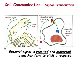
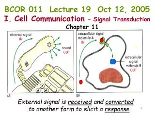
BCOR 011 Lecture 19 Oct 12, 2005 Cell Communication – Signal Transduction Chapter 11. External signal is received and converted to another form to elicit a response. Lecture Outline Types of intercellular communication The primary receiver – Receptors

E N D
BCOR 011 Lecture 19 Oct 12, 2005 • Cell Communication – Signal Transduction • Chapter 11 External signal is received and converted to another form to elicit a response
Lecture Outline • Types of intercellular communication • The primary receiver – Receptors • - the concept of AMPLIFICATION • Types of receptors • Ion Channels – Membrane depolarization • Trimeric G-Protein coupled receptors • - the cAMP signal pathway • - the phophatidyl inositol pathway, Ca++ release • Tyrosine Kinase – MAP Kinase Cascade • Internal cytosolic receptor systems
External signals are converted to Internal Responses • Cells sense and respond to the environment Prokaryotes: chemicals Humans: light - rods & cones of the eye sound – hair cells of inner ear chemicals in food – nose & tongue • Cells communicate with each other Direct contact Chemical signals
General principles: 1. Signals act over different ranges. 2. Signals have different chemical natures. 3. The same signal can induce a different response in different cells. 4. Cells respond to sets of signals. 5. Receptors relay signals via intracellular signaling cascades.
Signals act over different ranges Endocrine long distance ex. estrogen, epinephrine Paracrine local ex. nitric oxide, histamines, prostaglandins Like Fig 11.4 direct contact Cell-cell recognition ex. delta/notch Neuronal/Synaptic ex. neurotransmitters
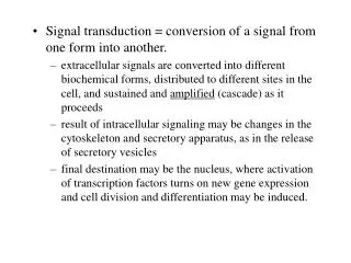
1º messenger Cells detect signal & respond Effector Enzymes Target Enzymes 2º messengers Signal transduction: ability of cell to translate receptor-ligand interaction into a change in behavior or gene expression
EXTRACELLULAR FLUID CYTOPLASM Plasma membrane 3 2 1 Reception Transduction Response Receptor Activation of cellular response Relay molecules in a signal transduction pathway Signal molecule Figure 11.5 Primary Messenger Target Enzymes Secondary Messengers Cascade Effect
- activates an enzyme activity, processes 100 substrates per second 1 primary signal Each protein in a signaling pathway • Amplifies the signal by activating multiple copies of the next component in the pathway Primary enzyme activates 100 target enzymes Each of the 100 enzymes activates an additional 100 dowstream target enzymes Each of the 10,000 downstream targets activates 100 control factors so rapidly have 1,000,000 active control factors
Receptors relay signals via intracellular SIGNALING CASCADES Push doorbell Ring bell Enzymatic activation of more ENZYMES amplification
Cell-surface receptors -large &/or hydrophilic ligands ion-channel-linked Trimeric G-protein-linked enzyme-linked (tyrosine kinase)
Gate closed Signalmolecule(ligand) Ions Ligand-gated ion channel receptor Plasma Membrane Gate open Cellularresponse Gate close Figure 11.7 Examples: Muscle Contraction Nerve Cell communication Ion channel receptors
Review: Remember the Na+/K+ ATPase (Na+/K+ pump)? [Na+] inside ~10mM; outside ~150mM [K+] inside ~100mM; outside ~5mM cell has membrane potential ~ -60mV Na+ Cl- -60mV K+ A- - + - - - + - + - + + +
Gated ion channels specifically let ions through membrane “keys”: small molecules (ligand-gated) or change in membrane potential (voltage-gated) + + + + + + + + + + + + + + + - - - - - - - - - - - - - - - -60 mV inside
Acetylcholine: common neurotransmitter opens ligand-gated Na+ channels on muscle cell and some nerve cells
Gated ion channels specifically let ions through membrane “keys”: small molecules (ligand-gated) + + + + + + + + + + + + + + + + - - - - - - - - - - - - - - - -60 mV inside
Gated ion channels specifically let ions through membrane “keys”: small molecules (ligand-gated) + + + + + + + + + + + + + + + + + + - - + - - - - - + - - - - + + - + + + + + +10 mV inside Influx of Na+ ions causes local, transient depolarization of membrane potential nerve impulse(action potential)
Gated ion channels specifically let ions through membrane “keys”: small molecules (ligand-gated) or change in membrane potential (voltage-gated) + + + + + + + + + + + + + + + + + + - - + - - - - + + - - - + + + + + + + + + + + + +10 mV inside Influx of Na+ ions causes local, transient depolarization of membrane potential nerve impulse(action potential)
Gated ion channels specifically let ions through membrane “keys”: small molecules (ligand-gated) or change in membrane potential (voltage-gated) + + + + + + + + + + + + + + + + + + + + - - - - - - - + + - - - - - + + + + + + + + + + + + +10 mV inside Influx of Na+ ions causes local, transient depolarization of membrane potential nerve impulse(action potential)
Gated ion channels specifically let ions through membrane “keys”: small molecules (ligand-gated) or change in membrane potential (voltage-gated) + + + + + + + + + + + + + + + + + + + + + - + - - - - - - + - - - - + + - - + + + - + + + + + + + +10 mV inside Influx of Na+ ions causes local, transient depolarization of membrane potential nerve impulse(action potential)
Transmission of action potential a. polarized b. Action potential Initiated by Ligand-gated Na+ channels opening Local depolarization Na+ Depolarization opens Voltage-gated Na+ channels Na+ K+ re-polarization Na+channels close K+ channels open Na+ K+ Action potential Propagates to as more Voltage-gated channels open
Action potential: nerve impulse; rapid, self-propagating electrical signal Muscle cell
Signal transmitted to muscle cell across a synapse Musclecell a. a. Depolarization opens voltage-gated Ca+2 channels Musclecell b. Ca+2 rushes in; Vesicles fuse with membrane b. c. Neurotransmitter released; opens ligand-gated Na+ channels on muscle cell Depolarizes muscle cell c. Signal: electrical to chemical to electrical
Depolarization of Muscle Cell Results in release of [Ca++]: Typically in cytosol Ca++ is 10-7 M, maintained ultra-low by active transport “pumps” Ca++/ATPases “vacuums” Ca++ Stored in Smooth Endoplasmic reticulum Ca++ from SER In Cytosol Triggers Activation of Myosin ATPase To “walk along” actin filaments – causing contraction
a. b. c. a,b Pesticides c Nerve gases Turning off the synapse…….. Acetylcholine degraded by acetylcholinesterase or removed by re-uptake & endocytosis if not removed…… Potent enzyme inhibitors Post-synaptic membrane can’t repolarize Paralysis, Tetany
Cell-surface receptors -large &/or hydrophilic ligands ion-channel-linked Trimeric G-protein-linked enzyme-linked (tyrosine kinase)
TrimericG protein-linked receptors: largest family of cell-surface receptors 7-pass membrane receptor ligand Ligand binding G-protein GTP activates G-protein by GTP exchange
Signal-binding site Segment that interacts with G proteins Inctivate enzyme ActivatedReceptor G-protein-linked Receptor Signal molecule Plasma Membrane GDP G-protein(inactive) GTP GDP CYTOPLASM Enzyme Activated Effector enzyme GTP GDP Pi Cellular response Figure 11.7 Trimeric G-protein-linked receptors
G-protein activation “molecular switch” inactive • (b) Ligand binds • G-protein associates • (c) GDP-GTP exchange • -Subunit dissociates Active G-Protein-GTP -> allosteric modulator of target effector enzyme active
All G-proteins – similar structure/activation • There are TWO broad subclasses of • trimeric G-protein-activated signal transduction pathways: • depends on theirtarget effector enzymes • A. adenylyl cyclase • B. phospholipase C
First messenger (signal molecule such as epinephrine) Adenylyl cyclase G protein GTP G-protein-linked receptor ATP cAMP Protein kinase A Cellular responses An activated Ga-protein-GTP • Can trigger the formation of cAMP, which then acts as a second messenger in cellular pathways Figure 11.10
G-protein-GTP activation of Effector Enzyme adenylylcyclase produces the 2nd messenger cAMP Activated G-protein Like Fig 11-9
cAMP activates target enzyme Protein Kinase A (PKA) Inactive PKA phosphorylates target proteins Active PKA
Phosphorylase kinase inactive + P active Protein Kinase A Phosphorylates downstream target enzymes Breaks down Starch Into Glucose
Reception Binding of epinephrine to G-protein-linked receptor (1 molecule) Transduction Inactive G protein Active G protein (102 molecules) Inactive adenylyl cyclase Active adenylyl cyclase (102) ATP Cyclic AMP (104) Inactive protein kinase A Active protein kinase A (104) Inactive phosphorylase kinase Active phosphorylase kinase (105) Inactive glycogen phosphorylase Active glycogen phosphorylase (106) Response Glycogen Glucose-1-phosphate(108 molecules) 1 A Signal Cascade amplification 102 104 105 106 108 Figure 11.13
What are targets for Protein Kinase A?? cAMP regulated pathways Function target tissue signal Glycogen breakdown muscle,liver epinephrine Heart rate cardiovascularepinephrine Water reabsorption kidney antidiuretic hormone
How to shut it off? No ligand G-protein -subunit is on a timer Inherent GTPase activity Auto Shut-off
How to shut it off? cAMP-phosphodiesterase rapidly cleaves cAMP (so short lived)
How do you turn it off? kinases – phosphatases Diametrically Opposed… Remember: whether you active or inactivate by adding P depends on the specific protein
What if you can’t turn off cascade? Vibrio cholera - causes cholera 7 great pandemics, Ganges Valley, Bangladesh Normal gut: H20, NaCl, NaHCO3 secretion controlled by hormones via Gs/cAMP signal pathways V. cholera – secretes enterotoxin, chemically modifies Gs – no GTPase activity - stays ON Severe watery diarrhea – dehydration, death
TWO subclasses of trimeric G-protein-activated signal transduction pathways: A. target protein adenylate cyclase cAMP-> PKA B. target protein phospholipase C
target effector enzyme is Phospholipase C PLC cleaves a membrane phospholipid (Phoshatidyl inositol) to two 2nd Messengers: Inositol-1,4,5-Trisphosphate (InsP3) & Diacylglycerol (DAG)
DAG Lipid Soluble InsP3 Water Soluble PIP2
DAG Activates Protein Kinase C (Starts Cascade) InsP3 Ligand for ER ligand- gated Ca++ channels Ca++ levels
Response: Protein Kinase C phosphorylates target proteins (ser & thr) cell growth regulation of ion channels cytoskeleton increases cell pH Protein secretion Ca++ Binds & activates calmodulin Calmodulin-binding proteins activated (kinases & phosphatases)
2 3 1 4 5 6 A signal molecule binds to a receptor, leading to activation of phospholipase C. DAG functions as a second messenger in other pathways. Phospholipase C cleaves a plasma membrane phospholipid called PIP2 into DAG and IP3. EXTRA- CELLULAR FLUID Signal molecule (first messenger) G protein DAG GTP PIP2 G-protein-linked receptor Phospholipase C IP3 (second messenger) IP3-gated calcium channel Endoplasmic reticulum (ER) Various proteins activated Cellularresponse Ca2+ Ca2+ (second messenger) The calcium ions activate the next protein in one or more signaling pathways. IP3 quickly diffuses through the cytosol and binds to an IP3– gated calcium channel in the ER membrane, causing it to open. Calcium ions flow out of the ER (down their con- centration gradient), raising the Ca2+ level in the cytosol. Figure 11.12
Summary • - signaling is endocrine, paracrine, synaptic, or direct cell contact • signal transduction is mediated by receptor proteins • Receptors bind primary signal (ligand) • Some amplification event occurs • Example: ligand gated ion channel opens • influx of ions triggers change in activity • (vesicle fusion in nerve end, contraction in muscle) • Example: ligand binds to 7-pass membrane receptor • catalyzes GTP exchange • to Ga-subunit of trimeric G-protein • active Ga-subunit-GTP is allosteric activator of • effector enzymes: • - ADENYLATE CYCLASE: makes cyclic AMP • - PHOSPHOLIPASE C: makes DAG and IP3 • these second messengers activate target enzymes • Trigger cascades • Must shut off cascade: removal of ligand, hydrolysis of GTP, • phosphodiesterase, protein phosphatases, Ca++ ion pumps